Edited, memorised or added to reading queue

on 20-Nov-2021 (Sat)

Do you want BuboFlash to help you learning these things? Click here to log in or create user.


#clinical #harrison #has-images #liver #medicine #surgery
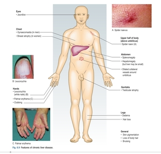
Typical physical findings in liver disease are icterus, hepatomegaly, hepatic tenderness, splenomegaly, spider angiomata, palmar erythema, and skin excoriations. Signs of advanced disease include muscle wast- ing, ascites, edema, dilated abdominal veins, hepatic fetor, asterixis, mental confusion, stupor, and coma. In male patients with cirrhosis, particularly that related to alcohol use, signs of hyperestrogenemia such as gynecomastia, testicular atrophy, and loss of male-pattern hair distribution may be found.
statusnot read reprioritisations
last reprioritisation on suggested re-reading day
started reading on finished reading on

pdf

cannot see any pdfs


#harrison #liver #medicine
Spider angiomata and palmar erythema occur in both acute and chronic liver disease
statusnot read reprioritisations
last reprioritisation on suggested re-reading day
started reading on finished reading on

pdf

cannot see any pdfs


#harrison #liver #medicine
Spider angiomata are superficial, tortuous arterioles, and—unlike simple telangiectases—typically fill from the center outward. Spider angiomata occur only on the arms, face, and upper torso; they can be pulsatile and may be difficult to detect in dark-skinned individuals.
statusnot read reprioritisations
last reprioritisation on suggested re-reading day
started reading on finished reading on

pdf

cannot see any pdfs


Flashcard 6977823837452

Tags
#clinical #harrison #has-images #liver #medicine #surgery


Question
Typical physical findings in liver disease are
Answer
icterus, hepatomegaly, hepatic tenderness, splenomegaly, spider angiomata, palmar erythema, and skin excoriations

statusnot learnedmeasured difficulty37% [default]last interval [days]               
repetition number in this series0memorised on               scheduled repetition               
scheduled repetition interval               last repetition or drill

Parent (intermediate) annotation

Open it
Typical physical findings in liver disease are icterus, hepatomegaly, hepatic tenderness, splenomegaly, spider angiomata, palmar erythema, and skin excoriations. Signs of advanced disease include muscle wast- ing, ascites, edema, di

Original toplevel document (pdf)

cannot see any pdfs


#Clinical #harrison #liver #medicine
Marked hepatomegaly is typical of cirrhosis, sinusoidal obstruction syndrome, infiltrative disorders such as amyloidosis, metastatic, or primary can- cers of the liver, and alcoholic hepatitis.
statusnot read reprioritisations
last reprioritisation on suggested re-reading day
started reading on finished reading on

pdf

cannot see any pdfs


#Clinical #harrison #liver #medicine
In patients with advanced liver disease, other factors frequently contribute to edema formation, including hypoalbuminemia, venous insufficiency, heart failure, and medications.
statusnot read reprioritisations
last reprioritisation on suggested re-reading day
started reading on finished reading on

pdf

cannot see any pdfs


#clinical #harrison #hepaticfaliure #liver #medicine
Hepatic failure is defined as the occurrence of signs or symptoms of hepatic encephalopathy in a person with severe acute or chronic liver disease.
statusnot read reprioritisations
last reprioritisation on suggested re-reading day
started reading on finished reading on

pdf

cannot see any pdfs


Flashcard 6977834847500

Tags
#clinical #harrison #hepaticfaliure #liver #medicine
Question
What is Hepatic Faliure?
Answer
Hepatic failure is defined as the occurrence of signs or symptoms of hepatic encephalopathy in a person with severe acute or chronic liver disease.

statusnot learnedmeasured difficulty37% [default]last interval [days]               
repetition number in this series0memorised on               scheduled repetition               
scheduled repetition interval               last repetition or drill

Parent (intermediate) annotation

Open it
Hepatic failure is defined as the occurrence of signs or symptoms of hepatic encephalopathy in a person with severe acute or chronic liver disease.

Original toplevel document (pdf)

cannot see any pdfs


hepatic enchephalopathy
#harrison #liver #medicine
The first signs of hepatic encephalopathy can be subtle and nonspecific—change in sleep patterns, change in personality, irritabil- ity, and mental dullness. Thereafter, confusion, disorientation, stupor, and eventually coma supervene. In acute liver failure, excitability and mania may be present. Physical findings include asterixis and flapping tremors of the body and tongue. Fetor hepaticus refers to the slightly sweet, ammoniacal odor that can develop in patients with liver failure, particularly if there is portal-venous shunting of blood around the liver. Other causes of coma and disorientation should be excluded, mainly electrolyte imbalances, sedative use, and renal or respiratory failure. The appearance of hepatic encephalopathy during acute hepatitis is the major criterion for diagnosis of fulminant hepatitis and indicates a poor prognosis. In chronic liver disease, encephalopathy is usually triggered by a medical complication such as gastrointestinal bleeding, over-diuresis, uremia, dehydration, electrolyte imbalance, infection, constipation, or use of narcotic analgesics.
statusnot read reprioritisations
last reprioritisation on suggested re-reading day
started reading on finished reading on

pdf

cannot see any pdfs


#harrison #liver #medicine
Other signs of advanced liver disease include umbilical hernia from ascites, hydrothorax, prominent veins over the abdomen, and caput medusa, a condition that consists of collateral veins radiating from the umbilicus and results from recanulation of the umbilical vein. Widened pulse pressure and signs of a hyperdynamic circulation can occur in patients with cirrhosis as a result of fluid and sodium retention, increased cardiac output, and reduced peripheral resistance. Patients with long-standing cirrhosis and portal hypertension are prone to develop the hepatopulmonary syndrome, which is defined by the triad of liver disease, hypoxemia, and pulmonary arteriovenous shunting. The hepatopulmonary syndrome is characterized by platypnea and orthodeoxia: shortness of breath and oxygen desaturation that occur paradoxically upon the assumption of an upright position. Measure- ment of oxygen saturation by pulse oximetry is a reliable screening test for hepatopulmonary syndrome
statusnot read reprioritisations
last reprioritisation on suggested re-reading day
started reading on finished reading on

pdf

cannot see any pdfs


Skin conditions in Liver Diseases
#harrison #liver #medicine
Hyperpigmentation is typical of advanced chronic cholestatic diseases such as primary biliary cholangitis and sclerosing cholangitis. In these same conditions, xanthelasma and tendon xanthomata occur as a result of retention and high serum levels of lipids and cholesterol. Slate-gray pigmentation of the skin is also seen with hemochromatosis if iron levels are high for a prolonged period. Mucocutaneous vasculitis with palpable purpura, especially on the lower extremities, is typical of cryoglobulinemia of chronic hepatitis C but can also occur in chronic hepatitis B.
statusnot read reprioritisations
last reprioritisation on suggested re-reading day
started reading on finished reading on

pdf

cannot see any pdfs


Specific physical signs of specific liver diseases
#harrison #liver #medicine
Some physical signs point to specific liver diseases. Kayser-Fleischer rings occur in Wilson disease and consist of a golden-brown copper pigment deposited in Descemet’s membrane at the periphery of the cornea; they are best seen by slit-lamp examination. Dupuytren con- tracture and parotid enlargement are suggestive of chronic alcoholism and alcoholic liver disease. In metastatic liver disease or primary hepatocellular carcinoma, signs of cachexia and wasting as well as firm hepatomegaly and a hepatic bruit may be prominent.
statusnot read reprioritisations
last reprioritisation on suggested re-reading day
started reading on finished reading on

pdf

cannot see any pdfs


#harrison #liver #medicine
The most common causes of acute liver disease are viral hepatitis (particularly hepatitis A, B, and C), drug-induced liver injury, cholangitis, and alcoholic liver disease.
statusnot read reprioritisations
last reprioritisation on suggested re-reading day
started reading on finished reading on

pdf

cannot see any pdfs


#harrison #liver #medicine
The most common causes of chronic liver disease, in general order of frequency, are chronic hepatitis C, alcoholic liver disease, nonalcoholic steatohepatitis, chronic hepatitis B, autoimmune hepatitis, sclerosing cholangitis, primary biliary cholangitis, hemochromatosis, and Wilson disease.
statusnot read reprioritisations
last reprioritisation on suggested re-reading day
started reading on finished reading on

pdf

cannot see any pdfs


#Pathology #cervix #obgyn
High-risk HPVs are by far the most important factor in the development of cervical cancer. HPVs are DNA viruses that are grouped into those of high and low oncogenic risk based on their genotypes. There are 15 high-risk HPVs that are currently identified, but HPV-16 alone accounts for almost 60% of cervical cancer cases, and HPV-18 accounts for another 10% of cases; other HPV types contribute to less than 5% of cases, individually. High-risk HPVs are also implicated in squamous cell carcinomas arising at many other sites, including the vagina, vulva, penis, anus, tonsil, and other oropharyngeal locations. As noted earlier, low oncogenic risk HPVs are the cause of sexually transmitted vulvar, perineal, and perianal warts (condyloma acuminatum).
statusnot read reprioritisations
last reprioritisation on suggested re-reading day
started reading on finished reading on

pdf

cannot see any pdfs


#Pathology #cervix #obgyn
Genital HPV infections are extremely common; most of them are asymptomatic, do not cause any tissue changes, and therefore are not detected on Pap test.
statusnot read reprioritisations
last reprioritisation on suggested re-reading day
started reading on finished reading on

pdf

cannot see any pdfs


#Pathology #cervix #obgyn
Productive, persistent HPV infection requires viral entry into immature basal epithelial cells. As a result, surfaces covered with mature, intact squamous epithelium, such as the ectocervix, vagina, vulva, penis, and oropharynx, are normally resistant to HPV infection. Sites in the female genital tract that are susceptible to infection include areas of squamous epithelial trauma and repair, where the virus may access basal cells, and the immature metaplastic squamous cells that are present at the squamocolumnar junction of the cervix (see Fig. 22.12). The cervix, with its relatively large areas of immature squamous metaplastic epithelium, is particularly vulnerable to HPV infection. Other sites in the body that are vulnerable to HPV infection include the squamocolumnar junction of the anus and the squamous cells of oropharyngeal tonsillar crypts, both relatively common sites of HPV-associated cancers in individuals who practice anal or oral sex, respectively.
statusnot read reprioritisations
last reprioritisation on suggested re-reading day
started reading on finished reading on

pdf

cannot see any pdfs


#Pathology #cervix #obgyn #virology
The ability of HPV to act as a carcinogen depends on the viral E6 and E7 proteins, which interfere with the activity of the key tumor suppressor proteins, p53 and RB, respectively.
statusnot read reprioritisations
last reprioritisation on suggested re-reading day
started reading on finished reading on

pdf

cannot see any pdfs


#Pathology #cervix #obgyn
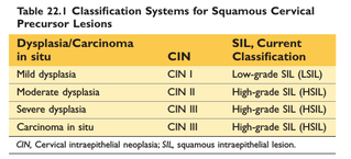
LSIL does not progress directly to invasive carcinoma, and, in fact, most cases regress spontaneously; only a small percentage progress to HSIL. LSIL represents a productive HPV infection in which there is a high level of viral replication, but only mild alterations in the growth of host cells. For these reasons, LSIL is not treated like a premalignant lesion. LSIL is approximately ten times more common than HSIL
statusnot read reprioritisations
last reprioritisation on suggested re-reading day
started reading on finished reading on

pdf

cannot see any pdfs


#Pathology
Classification of neplastic lessions of cervix
#Pathology #cervix #has-images #obgyn
statusnot read reprioritisations
last reprioritisation on suggested re-reading day
started reading on finished reading on

pdf

cannot see any pdfs


#Pathology #cervix #obgyn
The average age of patients with invasive cervical carcinoma is between 45 and 50 years. Squamous cell carcinoma is the most common histologic subtype, accounting for approxi- mately 80% of cases. The second most common tumor type is adenocarcinoma, which constitutes about 15% of cervical cancer cases and develops from a precursor lesion called adenocarcinoma in situ.
statusnot read reprioritisations
last reprioritisation on suggested re-reading day
started reading on finished reading on

pdf

cannot see any pdfs


#Pathology #cervix #histopathology #obgyn
Invasive cervical carcinoma may manifest as fungating (exophytic) or infiltrative masses. Squamous cell carcinoma is composed of nests and tongues of malignant squamous epithelium, either keratinizing or nonkeratinizing, which invade the underlying cervical stroma (Fig. 22.16A–B). Adenocarcinoma is characterized by proliferation of glandular epithelium composed of malignant endocervical cells with large, hyperchromatic nuclei and relatively mucin-depleted cytoplasm, resulting in a dark appearance of the glands, as compared with the normal endocervical epithelium
statusnot read reprioritisations
last reprioritisation on suggested re-reading day
started reading on finished reading on

pdf

cannot see any pdfs


metastases of Advance Ca cervix
#Pathology #cervix #obgyn
Advanced cervical carcinoma spreads by direct exten- sion to contiguous tissues, including paracervical soft tissue, urinary bladder, ureters (resulting in hydronephrosis), rectum, and vagina. Lymphovascular invasion results in local and distant lymph nodes metastases. Distant metastases may also be found in the liver, lungs, bone marrow, and other organs.
statusnot read reprioritisations
last reprioritisation on suggested re-reading day
started reading on finished reading on

pdf

cannot see any pdfs


pap smear
#Pathology #cervix #has-images #obgyn
The reason that cytologic screening is so effective in preventing cervical cancer is that most cancers arise from precursor lesions over the course of years. These lesions shed abnormal cells that can be detected on cytologic examination. Using a spatula or brush, the transformation zone of the cervix is circumferentially scraped and the cells are smeared or spun down onto a slide. Following fixation and staining with the Papanicolaou method, the smears are screened microscopically by eye or (increasingly) with automated image analysis systems.
statusnot read reprioritisations
last reprioritisation on suggested re-reading day
started reading on finished reading on

pdf

cannot see any pdfs


#Pathology #mensturation #obgyn #physiology
The endometrium undergoes dynamic physiologic and morphologic changes during the menstrual cycle in response to sex steroid hormones coordinately produced in the ovary.
statusnot read reprioritisations
last reprioritisation on suggested re-reading day
started reading on finished reading on

pdf

cannot see any pdfs


#Pathology #menstruation #obgyn #physiology
The histologic appearance of the endometrium may be used to assess hormonal status, document ovulation, and determine causes of endometrial bleeding and infertility
statusnot read reprioritisations
last reprioritisation on suggested re-reading day
started reading on finished reading on

pdf

cannot see any pdfs


#Pathology
• The cycle commences with menses, during which the superficial portion of the endometrium, referred to as the functionalis, is shed. • The proliferative phase is marked by rapid growth of glands and stroma arising from the deeper portion of the endometrium (basalis). During the proliferative phase, the glands are straight, tubular structures lined by regular, tall, pseudostratified columnar cells. Mitotic figures are numerous, and there is no evidence of mucus secretion or vacuolation. The endometrial stroma is composed of spindle cells with scant cytoplasm that are also actively proliferating (see Fig. 22.19A). • At ovulation, endometrial proliferation ceases and dif- ferentiation commences in response to the effects of progesterone made by the corpus luteum in the ovary. • Postovulation is initially marked by the appearance of secretory vacuoles beneath the nuclei in the glandular epithelium (see Fig. 22.19B). Secretory activity is most prominent during the third week of the menstrual cycle, when the basal vacuoles progressively move to the apical surface. By the fourth week, the glands are tortuous, producing a serrated appearance. This serrated or “sawtooth” appearance is accentuated by secretory exhaustion and shrinkage of the glands. • Stromal changes in the late secretory phase, due predomi- nantly to progesterone, are the most significant features. Prominent spiral arterioles appear accompanied by an
statusnot read reprioritisations
last reprioritisation on suggested re-reading day
started reading on finished reading on

pdf

cannot see any pdfs


#Pathology
increase in ground substance and edema between the stromal cells followed by stromal cell hypertrophy, increased cytoplasmic eosinophilia (predecidual change), and a resurgence of stromal mitoses (see Fig. 22.19C). Predecidual changes spread throughout the functio- nalis and are accompanied by a sparse infiltrate of neutrophils and lymphocytes, which in this context are considered normal. • With the dissolution of the corpus luteum and the subsequent drop in progesterone levels, the functionalis degenerates and bleeding into the stroma occurs, followed by stromal breakdown and onset of the next menstrual cycle
statusnot read reprioritisations
last reprioritisation on suggested re-reading day
started reading on finished reading on

pdf

cannot see any pdfs


#Pathology
1002 CHAPTER 22 The Female Genital Tract down-regulates the expression of estrogen receptor in both the glands and the stroma, and as a result endometrial proliferation is suppressed. Progesterone also promotes the differentiation of the glands and causes functional changes in the stromal cells. Endometrial stem cells have been identi- fied that likely have a central role in the regeneration of the endometrium after menses. They may also contribute to the development of ectopic endometrial tissue and endo- metrial cancer. FUNCTIONAL ENDOMETRIAL DISORDERS (DYSFUNCTIONAL UTERINE BLEEDING) Although abnormal uterine bleeding can be caused by well-defined pathologic conditions, such as chronic endo- metritis, endometrial polyps (Fig. 22.20C), submucosal leiomyomas (see Fig. 22.20D), or endometrial neoplasms, it most commonly stems from hormonal disturbances that produce dysfunctional uterine bleeding (Table 22.3). This is a clinical term for uterine bleeding that lacks an underlying structural abnormality. As discussed earlier, the normal increase in ground substance and edema between the stromal cells followed by stromal cell hypertrophy, increased cytoplasmic eosinophilia (predecidual change), and a resurgence of stromal mitoses (see Fig. 22.19C). Predecidual changes spread throughout the functio- nalis and are accompanied by a sparse infiltrate of neutrophils and lymphocytes, which in this context are considered normal. • With the dissolution of the corpus luteum and the subsequent drop in progesterone levels, the functionalis degenerates and bleeding into the stroma occurs, followed by stromal breakdown and onset of the next menstrual cycle
statusnot read reprioritisations
last reprioritisation on suggested re-reading day
started reading on finished reading on

pdf

cannot see any pdfs