Edited, memorised or added to reading queue

on 24-Sep-2021 (Fri)

Do you want BuboFlash to help you learning these things? Click here to log in or create user.

Seizures and epilepsy
#Neurology
A seizure (from the Latin sacire, “to take possession of”) is a transient occurrence of signs or symptoms due to abnormal excessive or syn- chronous neuronal activity in the brain. Depending on the distribution of discharges, this abnormal brain activity can have various mani- festations, ranging from dramatic convulsive activity to experiential phenomena not readily discernible by an observer. Although a variety of factors influence the incidence and prevalence of seizures, ~5–10% of the population will have at least one seizure, with the highest incidence occurring in early childhood and late adulthood. The meaning of the term seizure needs to be carefully distinguished from that of epilepsy. Epilepsy describes a condition in which a person has a risk of recurrent seizures due to a chronic, underlying process. This definition implies that a person with a single seizure, or recur- rent seizures due to correctable or avoidable circumstances, does not necessarily have epilepsy (although a single seizure associated with particular clinical or electroencephalographic features may establish the diagnosis of epilepsy). Epilepsy refers to a clinical phenomenon rather than a single disease entity, because there are many forms and causes of epilepsy. However, among the many causes of epilepsy there are various epilepsy syndromes in which the clinical and pathologic char- acteristics are distinctive and suggest a specific underlying etiology
statusnot read reprioritisations
last reprioritisation on suggested re-reading day
started reading on finished reading on

pdf

cannot see any pdfs


Seizures classification
#Neurology

A fundamental principle is that seizures may be either focal or gen- eralized. Focal seizures originate within networks limited to one brain region (note that the term partial seizures is no longer used). Generalized

statusnot read reprioritisations
last reprioritisation on suggested re-reading day
started reading on finished reading on

pdf

cannot see any pdfs


seizures classification 2
#Neurology
seizures arise within and rapidly engage networks distributed across both cerebral hemispheres. Focal seizures are usually associated with structural abnormalities of the brain. In contrast, generalized seizures may result from cellular, biochemical, or structural abnormalities that have a more widespread distribution. There are clear exceptions in both cases, however.
statusnot read reprioritisations
last reprioritisation on suggested re-reading day
started reading on finished reading on

pdf

cannot see any pdfs


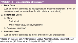
classification of seizures
#Neurology #has-images
statusnot read reprioritisations
last reprioritisation on suggested re-reading day
started reading on finished reading on

pdf

cannot see any pdfs


Focal seizures arise from a neuronal network either discretely localized within one brain region or more broadly distributed but still within a cerebral hemisphere
statusnot read reprioritisations
last reprioritisation on suggested re-reading day
started reading on finished reading on

pdf

cannot see any pdfs


Focal seizures can also evolve into generalized seizures. In the past this was referred to as focal seizures with secondary generalization,
statusnot read reprioritisations
last reprioritisation on suggested re-reading day
started reading on finished reading on

pdf

cannot see any pdfs


The routine interictal (i.e., between seizures) electroencephalogram (EEG) in patients with focal seizures is often normal or may show brief discharges termed epileptiform spikes, or sharp waves.
statusnot read reprioritisations
last reprioritisation on suggested re-reading day
started reading on finished reading on

pdf

cannot see any pdfs


Focal seizures can have motor manifestations (such as tonic, clonic, or myoclonic movements) or nonmotor manifestations (such as sensory, autonomic, or emotional symptoms) without impairment of awareness.
statusnot read reprioritisations
last reprioritisation on suggested re-reading day
started reading on finished reading on

pdf

cannot see any pdfs


The EEG recorded with scalp electrodes during the seizure (i.e., an ictal EEG) may show abnormal discharges in a very limited region over the appropriate area of cerebral cortex if the seizure focus involves the cerebral convexity.
statusnot read reprioritisations
last reprioritisation on suggested re-reading day
started reading on finished reading on

pdf

cannot see any pdfs


Three additional features of focal motor seizures are worth noting. First, in some patients, the abnormal motor movements may begin in a very restricted region such as the fingers and gradually progress (over seconds to minutes) to include a larger portion of the extremity. This phenomenon, described by Hughlings Jackson and known as a “Jacksonian march,” represents the spread of seizure activity over a progressively larger region of motor cortex. Second, patients may expe- rience a localized paresis (Todd’s paralysis) for minutes to many hours in the involved region following the seizure. Third, in rare instances, the seizure may continue for hours or days. This condition, termed epilepsia partialis continua, is often refractory to medical therapy
statusnot read reprioritisations
last reprioritisation on suggested re-reading day
started reading on finished reading on

pdf

cannot see any pdfs


#Neurology
Focal seizures may also manifest as changes in somatic sensation (e.g., paresthesias), vision (flashing lights or formed hallucinations), equilibrium (sensation of falling or vertigo), or autonomic function (flushing, sweating, piloerection)
statusnot read reprioritisations
last reprioritisation on suggested re-reading day
started reading on finished reading on

pdf

cannot see any pdfs


Some patients describe odd, internal feelings such as fear, a sense of impending change, detachment, depersonalization, déjá vu, or illusions that objects are growing smaller (micropsia) or larger (macropsia). These subjective, “internal” events that are not directly observable by someone else are referred to as auras.
statusnot read reprioritisations
last reprioritisation on suggested re-reading day
started reading on finished reading on

pdf

cannot see any pdfs


Focal seizures may also be accompanied by a transient impairment of the patient’s ability to maintain normal contact with the environment. The patient is unable to respond appropriately to visual or verbal commands during the seizure and has impaired recollection or awareness of the ictal phase. The seizures frequently begin with an aura (i.e., a focal sei- zure without cognitive disturbance) that is stereotypic for the patient. The start of the ictal phase is often a motionless stare, which marks the onset of the period of impaired awareness. The impaired awareness is usually accompanied by automatisms, which are involuntary, automatic behaviors that have a wide range of manifestations. Automatisms may consist of very basic behaviors such as chewing, lip smacking, swallowing, or “picking” movements of the hands, or more elabo- rate behaviors such as a display of emotion or running. The patient is typically confused following the seizure, and the transition to full recovery of consciousness may range from seconds up to an hour or longer. Examination immediately following the seizure may show an anterograde amnesia or transient neurological deficits (such as aphasia, hemi-neglect, or visual loss) caused by postictal inhibition of the corti- cal regions most involved in the seizure itself.
statusnot read reprioritisations
last reprioritisation on suggested re-reading day
started reading on finished reading on

pdf

cannot see any pdfs


Focal seizures can spread to involve both cerebral hemispheres and produce a generalized seizure, usually of the tonic-clonic variety (discussed below). This evolution is observed frequently following focal seizures arising from a region in the frontal lobe, but may also be associated with focal seizures occurring elsewhere in the brain. A focal seizure that evolves into a generalized seizure is often difficult to distinguish from a primary generalized onset tonic-clonic seizure, because bystanders tend to emphasize the more dramatic, generalized convulsive phase of the seizure and overlook the more subtle, focal symptoms present at onset. In some cases, the focal onset of the seizure becomes apparent only when a careful history identifies a preceding aura. Often, however, the focal onset is not clinically evident and may be established only through careful EEG analysis. Nonetheless, distin- guishing between these two entities is extremely important, because there may be substantial differences in the evaluation and treatment of epilepsies characterized by focal versus generalized onset seizures
statusnot read reprioritisations
last reprioritisation on suggested re-reading day
started reading on finished reading on

pdf

cannot see any pdfs


Typical absence seizures are charac- terized by sudden, brief lapses of consciousness without loss of pos- tural control. The seizure usually lasts for only seconds, consciousness returns as suddenly as it was lost, and there is no postictal confusion. Although the brief loss of consciousness may be clinically inapparent or the sole manifestation of the seizure discharge, absence seizures are usually accompanied by subtle, bilateral motor signs such as rapid blinking of the eyelids, chewing movements, or small-amplitude, clonic movements of the hands.
statusnot read reprioritisations
last reprioritisation on suggested re-reading day
started reading on finished reading on

pdf

cannot see any pdfs


Typical absence seizures are associated with a group of genetically determined epilepsies with onset usually in childhood (ages 4–10 years) or early adolescence and are the main seizure type in 15–20% of chil- dren with epilepsy.
statusnot read reprioritisations
last reprioritisation on suggested re-reading day
started reading on finished reading on

pdf

cannot see any pdfs


The electrophysiologic hallmark of typical absence seizures is a generalized, symmetric, 3-Hz spike-and-slow-wave discharges that begins and ends suddenly, superimposed on a normal EEG back- ground.
statusnot read reprioritisations
last reprioritisation on suggested re-reading day
started reading on finished reading on

pdf

cannot see any pdfs


Atypical absence seizures have fea- tures that deviate both clinically and electrophysiologically from typi- cal absence seizures. For example, the lapse of consciousness is usually of longer duration and less abrupt in onset and cessation, and the seizure is accompanied by more obvious motor signs that may include focal or lateralizing features. The EEG shows a generalized, slow spike- and-slow-wave pattern with a frequency of ≤2.5 per second, as well as other abnormal activity. Atypical absence seizures are usually associ- ated with diffuse or multifocal structural abnormalities of the brain and therefore may accompany other signs of neurologic dysfunction such as mental retardation. Furthermore, the seizures are less responsive to anticonvulsants compared to typical absence seizures.
statusnot read reprioritisations
last reprioritisation on suggested re-reading day
started reading on finished reading on

pdf

cannot see any pdfs


They are also the most common seizure type resulting from metabolic derangements and are therefore frequently encountered in many different clinical settings. The seizure usually begins abruptly without warning, although some patients describe vague premonitory symptoms in the hours leading up to the seizure. This prodrome is distinct from the stereotypic auras associated with focal seizures that generalize. The initial phase of the seizure is usually tonic contraction of muscles throughout the body, accounting for a number of the classic features of the event. Tonic contraction of the muscles of expiration and the larynx at the onset will produce a loud moan or “ictal cry.” Respirations are impaired, secretions pool in the oropharynx, and cya- nosis develops. Contraction of the jaw muscles may cause biting of the tongue. A marked enhancement of sympathetic tone leads to increases in heart rate, blood pressure, and pupillary size. After 10–20 s, the tonic phase of the seizure typically evolves into the clonic phase, produced by the superimposition of periods of muscle relaxation on the tonic muscle contraction. The periods of relaxation progressively increase until the end of the ictal phase, which usually lasts no more than 1 min. The pos- tictal phase is characterized by unresponsiveness, muscular flaccidity, and excessive salivation that can cause stridorous breathing and partial airway obstruction. Bladder or bowel incontinence may occur at this point. Patients gradually regain consciousness over minutes to hours, and during this transition, there is typically a period of postictal confu- sion. Patients subsequently complain of headache, fatigue, and muscle ache that can last for many hours. The duration of impaired conscious- ness in the postictal phase can be extremely long (i.e., many hours) in patients with prolonged seizures or underlying central nervous system (CNS) diseases such as alcoholic cerebral atrophy
statusnot read reprioritisations
last reprioritisation on suggested re-reading day
started reading on finished reading on

pdf

cannot see any pdfs


Brief tonic seizures last- ing only a few seconds are especially noteworthy since they are usually associated with specific epilepsy syndromes having mixed seizure phenotypes, such as the Lennox-Gastaut syndrome
statusnot read reprioritisations
last reprioritisation on suggested re-reading day
started reading on finished reading on

pdf

cannot see any pdfs


2 • Higher the shape factor, more ductile will be the section and will give greater deflection at collapse
statusnot read reprioritisations
last reprioritisation on suggested re-reading day
started reading on finished reading on

pdf

cannot see any pdfs


Flashcard 6770921704716

Question
[default - edit me]
Answer
de! f:gh?>g;':i>j ki=lhmn':i>j H opp M ! q:>rghs'?i?;mg's>t>shgu v>w;'?i?;mg's>t>shgu H xyzz{ M

statusnot learnedmeasured difficulty37% [default]last interval [days]               
repetition number in this series0memorised on               scheduled repetition               
scheduled repetition interval               last repetition or drill

pdf

cannot see any pdfs


Fouierners gangrene
#scrotum
Fournier’s gangrene is an uncommon and nasty condition (Figure 80.21) characterised by a polymicrobial infection of the soft tissues of the perineum, external genitalia and peri- anal region. It is a form of necrotising fasciitis. There is rapid onset of gangrene leading to exposure of the scrotal contents. Although it can occur in conjunction with sepsis of the tes- tis, epididymis or perianal region, an obvious cause is absent in over half the cases. It can arise following minor injuries or procedures in the perineal area, such as a bruise, scratch, urethral dilatation, injection of haemorrhoids or opening of a periurethral abscess. Many patients have concurrent illnesses that diminish their defences, most notably diabetes mellitus and alcoholism. There is a mixed infection of aerobic and anaerobic bacte- ria in a fulminating inflammation of the subcutaneous tissues, which results in an obliterative arteritis of the arterioles to the scrotal skin that in turn results in gangrene. The condition can spread rapidly to involve the fascia and skin of the penis, perineum and abdominal wall
statusnot read reprioritisations
last reprioritisation on suggested re-reading day
started reading on finished reading on

pdf

cannot see any pdfs


CHAPTER 80 Testis and scrotum PART 12 | GENITOURINARY 1510 Clinical features There is sudden pain in the scrotum associated with pros- tration, pallor and pyrexia. Cellulitis spreads rapidly (within hours) with small necrotic areas of skin which, if untreated, coalesce to involve the entire scrotal and penile coverings, which may then slough, leaving the testes exposed but healthy. There may be crepitus and a foul-smelling exudate. The patient typically becomes septic and severely unwell in a short period of time
statusnot read reprioritisations
last reprioritisation on suggested re-reading day
started reading on finished reading on

pdf

cannot see any pdfs


Treatment of a case of Fournier’s gangrene is a surgical emer- gency. Initial management involves intravenous fluid resusci- tation and early use of broad spectrum intravenous antibiotics. Urgent wide surgical excision of the dead and infected tissue is essential and the extent of the internal necrosis is typically much greater than the external appearances suggest, such that extensive debridement is often necessary. Urinary and faecal diversion may be necessary. Supportive care is essential, because the patients often become severely septic. Early review of the wounds is helpful to confirm that all dead tissue has been removed, and when the infection has been controlled, vacuum-assisted dressing is helpful, if it is available. If the patient survives the acute episode, skin graft- ing is often necessary. Despite best therapy, mortality rates as high as 50% are often reported
statusnot read reprioritisations
last reprioritisation on suggested re-reading day
started reading on finished reading on

pdf

cannot see any pdfs


A hydrocoele is an abnormal collection of serous fluid in a part of the processus vaginalis, usually the tunica vaginalis
statusnot read reprioritisations
last reprioritisation on suggested re-reading day
started reading on finished reading on

pdf

cannot see any pdfs


A hydrocoele can be produced in four different ways (Figure 80.9): 1 By connection with the peritoneal cavity via a patent pro- cessus vaginalis (congenital). 2 By excessive production of fluid within the sac, e.g. a sec- ondary hydrocoele.
statusnot read reprioritisations
last reprioritisation on suggested re-reading day
started reading on finished reading on

pdf

cannot see any pdfs


By defective absorption of fluid; this appears to be the explanation for most primary hydrocoeles, although the reason why the fluid is not absorbed is obscure. They are sometimes called vaginal hydrocoeles. 4 By interference with the lymphatic drainage of scrotal structures
statusnot read reprioritisations
last reprioritisation on suggested re-reading day
started reading on finished reading on

pdf

cannot see any pdfs


Secondary hydrocele
#Surgery
A secondary hydrocoele is most frequently associated with acute or chronic epididymo-orchitis. It is also seen with torsion of the testis and with some testicular tumours. A secondary hydrocoele is usually lax and is typically small: the underlying testis is usually palpable. If a tumour is sus- pected, the hydrocoele should not be punctured for fear of needle-track implantation of malignant cells. A secondary hydrocoele subsides when the primary lesion resolves
statusnot read reprioritisations
last reprioritisation on suggested re-reading day
started reading on finished reading on

pdf

cannot see any pdfs