Edited, memorised or added to reading queue

on 02-Dec-2021 (Thu)

Do you want BuboFlash to help you learning these things? Click here to log in or create user.

Fracture healing 1
#fracture #orthopedics
The healing of fractures is in many ways similar to the healing of soft tissue wounds, except that soft tissue heals with fibrous tissue, and end result of bone healing is mineralised mesenchymal tissue, i.e. bone. A fracture begins to heal soon after it occurs, through a continuous series of stages
statusnot read reprioritisations
last reprioritisation on suggested re-reading day
started reading on finished reading on

pdf

cannot see any pdfs


Flashcard 6989680610572

Tags
#fracture #orthopedics
Question
STAGES IN FRACTURE HEALING OF CORTICAL BONE
Answer
• Stage of haematoma • Stage of granulation tissue • Stage of callus • Stage of r emodelling (formerly called consolidation) • Stage of modelling (formerly called r emodelling)

statusnot learnedmeasured difficulty37% [default]last interval [days]               
repetition number in this series0memorised on               scheduled repetition               
scheduled repetition interval               last repetition or drill

pdf

cannot see any pdfs


Stage of Haematoma
#fracture #orthopedics
This stage lasts up to 7 days. When a bone is fractured, blood leaks out through torn vessels in the bone and forms a haematoma between and around the fracture. The periosteum and local soft tissues are stripped from the fracture ends. This results in ischaemic necrosis of the fracture ends over a variable length, usually only a few millimetres. Deprived of their blood supply, some osteocytes die whereas others are sensitised to respond subsequently by differentiating into daughter cells. These cells later contribute to the healing process.
statusnot read reprioritisations
last reprioritisation on suggested re-reading day
started reading on finished reading on

pdf

cannot see any pdfs


Stage of Callus
#fracture #orthopedics
This stage lasts for about 4-12 weeks. In this stage, the granulation tissue differentiates further and creates osteoblasts. These cells lay down an intercellular matrix which soon becomes impregnated with calcium salts. This results in formulation of the callus, also called woven bone. The callus is the first sign of union visible on X-rays, usually 3 weeks after the fracture (Fig-2.4). The formation of this bridge of woven bone imparts good strength to the fracture. Callus formation is slower in adults than in children, and in cortical bones than in cancellous bones
statusnot read reprioritisations
last reprioritisation on suggested re-reading day
started reading on finished reading on

pdf

cannot see any pdfs


Stage of remodelling
#fracture #orthopedics
Formerly called the stage of consolidation. In this stage, the woven bone is replaced by mature bone with a typical lamellar structure. This process of change is multicellular unit based, whereby a pocket of callus is replaced by a pocket of lamellar bone. It is a slow process and takes anything from one to four years.
statusnot read reprioritisations
last reprioritisation on suggested re-reading day
started reading on finished reading on

pdf

cannot see any pdfs


Stage of modelling
#fracture #orthopedics
Formerly called the stage of remodelling. In this stage the bone is gradually strengthened. The shapening of cortices occurs at the endosteal and periosteal surfaces. The major stimulus to this process comes from local bone strains i.e., weight bearing stresses and muscle forces when the person resumes activity. This stage is more conspicuous in children with angulated fractures. It occurs to a very limited extent in fractures in adults.
statusnot read reprioritisations
last reprioritisation on suggested re-reading day
started reading on finished reading on

pdf

cannot see any pdfs


Healing of Cancellous bone
#fracture #orthopedics
The healing of fractured cancellous bone follows a different pattern. The bone is of uniform spongy texture and has no medullary cavity so that there is a large area of contact between the trabeculae. Union can occur directly between the bony trabeculae. Subsequent to haematoma and granulation formation, mature osteoblasts lay down woven bone in the intercellular matrix, and the two fragments unite.
statusnot read reprioritisations
last reprioritisation on suggested re-reading day
started reading on finished reading on

pdf

cannot see any pdfs


Flashcard 6989694766348

Tags
#fracture #orthopedics
Question
Most common cause of non-union is [...] .
Answer
Most common cause of non-union is inadequate immobilisation.

statusnot learnedmeasured difficulty37% [default]last interval [days]               
repetition number in this series0memorised on               scheduled repetition               
scheduled repetition interval               last repetition or drill

pdf

cannot see any pdfs


Flashcard 6989697125644

Tags
#fracture #orthopedics
Question
Pathognomonic sign of traumatic and fresh fracture is [...] .
Answer
P athognomonic sign of traumatic and fresh fracture is crepitus.

statusnot learnedmeasured difficulty37% [default]last interval [days]               
repetition number in this series0memorised on               scheduled repetition               
scheduled repetition interval               last repetition or drill

pdf

cannot see any pdfs


#Surgery
#Surgery #has-images #wound
statusnot read reprioritisations
last reprioritisation on suggested re-reading day
started reading on finished reading on

pdf

cannot see any pdfs


Flashcard 6989702892812

Tags
#Pathology #surgery #wound
Question
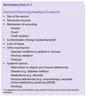
Glucocorticoids (steroids) have well-documented antiinflam- matory effects, and their administration may result in weakness of the scar due to [...] production and [...]
Answer
Glucocorticoids (steroids) have well-documented antiinflam - matory effects, and their administration may result in weakness of the scar due to inhibition of TGF- β production and diminished fibrosis.

statusnot learnedmeasured difficulty37% [default]last interval [days]               
repetition number in this series0memorised on               scheduled repetition               
scheduled repetition interval               last repetition or drill

pdf

cannot see any pdfs


#Surgery
#Surgery #has-images #wound
statusnot read reprioritisations
last reprioritisation on suggested re-reading day
started reading on finished reading on

pdf

cannot see any pdfs


Flashcard 6989708659980

Tags
#compartment #fracture #orthopedics
Question
COMPARTMENT SYNDROME
Answer
The limbs contain muscles in compartments enclosed by bones, fascia and interosseous membrane (Fig-7.1). A rise in pressure within compartments due to any reason may jeopar dize the blood supply to the muscles and nerves withi n the compartment, resulting in what is known as “compartment syndrome”.

statusnot learnedmeasured difficulty37% [default]last interval [days]               
repetition number in this series0memorised on               scheduled repetition               
scheduled repetition interval               last repetition or drill

pdf

cannot see any pdfs


Flashcard 6989711805708

Tags
#compartment #fracture #fracturecomplications #orthopedics
Question
Injuries with a high risk of developing compartment syndrome are as follows:
Answer
• Supracondylar fracture of the humerus • Forearm bone fractures • Closed tibial fractures • Crush injuries to leg and forearm.

statusnot learnedmeasured difficulty37% [default]last interval [days]               
repetition number in this series0memorised on               scheduled repetition               
scheduled repetition interval               last repetition or drill

pdf

cannot see any pdfs


Diagnosis of compartment syndrome
#compartment #fracture #fracturecomplications #orthopedics
Compartment syndrome can be diagnosed early by high index of suspicion. Excessive pain, not relieved with usual doses of analgesics, in a patient with an injury known to cause compartment syndrome must raise an alarm in the mind of the treating doctor. Injuries with a high risk of developing compartment syndrome are as follows: • Supracondylar fracture of the humerus • Forearm bone fractures • Closed tibial fractures • Crush injuries to leg and forearm. Stretch test: This is the earliest sign of impending compartment syndrome. The ischaemic muscles, when stretched, give rise to pain. It is possible to stretch the affected muscles by passively moving the joints in direction opposite to that of the damaged muscle’s action. (e.g., passive extension of fingers produces pain in flexor compartment of the forearm). Other signs include a tense compartment, hypo- aesthesia in the distribution of involved nerves, muscle weakness etc. Compartment syndrome can be confirmed by measuring compartment pressure. A pressure higher than 40 mm of water is indica- tive of compartment syndrome. Pulses may remain palpable till very late in impending compartment syndrome, and should not provide a false sense of security that all is well.
statusnot read reprioritisations
last reprioritisation on suggested re-reading day
started reading on finished reading on

pdf

cannot see any pdfs


#orthopedics
Eaton and greene cycle for compartment syndrome development
#compartment #fracture #fracturecomplications #has-images #orthopedics
statusnot read reprioritisations
last reprioritisation on suggested re-reading day
started reading on finished reading on

pdf

cannot see any pdfs


Flashcard 6989723340044

Tags
#compartment #fracture #fracturecomplications #orthopedics
Question
t/t for compartment syndrome
Answer

Preventive measures:-

  1. proper montoring of the injuries that are at ahigh risk of developing compartment syndrome.
  2. limb elevation
  3. activer finger movements

defenitive:- to prevent complications

Early surgical decompression

Fasciotomy: The deep fascia of the compartment is slit longitudinally (e.g., in forearm).

Fibulectomy: The middle third of the fibula is excised in order to decompress all compart ments of the leg.


statusnot learnedmeasured difficulty37% [default]last interval [days]               
repetition number in this series0memorised on               scheduled repetition               
scheduled repetition interval               last repetition or drill

pdf

cannot see any pdfs


Delayed union and Non union
#fracture #fracturecomplications #orthopedics
When a fracture takes more than the usual time to unite, it is said to have gone in delayed union. A large percentage of such fractures eventually unite. In some, the union does not progress, and they fail to unite. These are called non-union. Conventionally, it is not before 6 months that a fracture can be declared as non-union. It is often difficult to say whether the fracture is in delayed union, or has gone into non- union. Only progressive evaluation of the X-rays over a period of time can solve this issue. Presence of mobility at the fracture after a reasonable period is surely a sign of non-union. Presence of pain at the fracture site on using the limbs also indicates non-union. Non-union may be painless if pseudo joint forms between the fracture ends (pseudoarthrosis).
statusnot read reprioritisations
last reprioritisation on suggested re-reading day
started reading on finished reading on

pdf

cannot see any pdfs


Flashcard 6989727534348

Tags
#fracture #fracturecomplications #orthopedics
Question
Sites where non-union occurs commonly are
Answer
neck of the femur , scaphoid, lower third of the tibia, lower thir d of the ulna and lateral condyle of the humerus.

statusnot learnedmeasured difficulty37% [default]last interval [days]               
repetition number in this series0memorised on               scheduled repetition               
scheduled repetition interval               last repetition or drill

pdf

cannot see any pdfs


Pathogenesis of heart failure with a depressed ejection fraction 2
#CVS #HF #harrison #medicine
LV remodeling devel- ops in response to a series of complex events that occur at the cellular and molecular levels (Table 252-3). These changes include (1) myocyte hypertrophy; (2) alterations in the contractile properties of the myo- cyte; (3) progressive loss of myocytes through necrosis, apoptosis, and autophagic cell death; (4) β-adrenergic desensitization; (5) abnormal myocardial energetics and metabolism; and (6) reorganization of the extracellular matrix with dissolution of the organized structural col- lagen weave surrounding myocytes and subsequent replacement by an interstitial collagen matrix that does not provide structural support to the myocytes. The biologic stimuli for these profound changes include mechanical stretch of the myocyte, circulating neurohormones (e.g., norepinephrine, angiotensin II), inflammatory cytokines (e.g., tumor necrosis factor [TNF]), other peptides and growth factors (e.g., endo- thelin), and reactive oxygen species (e.g., superoxide). The sustained overexpression of these biologically active molecules contributes to the progression of HF by virtue of the deleterious effects they exert on the heart and the circulation. Indeed, this insight forms the clinical rationale for using pharmacologic agents that antagonize these systems (e.g., angiotensin-converting enzyme [ACE] inhibitors, angiotensin receptor-neprilysin inhibitors [ARNIs] and beta blockers) in treating patients with HF
statusnot read reprioritisations
last reprioritisation on suggested re-reading day
started reading on finished reading on

pdf

cannot see any pdfs