Edited, memorised or added to reading queue
on 10-Dec-2018 (Mon)
Do you want BuboFlash to help you learning these things? Click here to log in or create user.
Flashcard 3628064181516
| status | not learned | measured difficulty | 37% [default] | last interval [days] | |||
|---|---|---|---|---|---|---|---|
| repetition number in this series | 0 | memorised on | scheduled repetition | ||||
| scheduled repetition interval | last repetition or drill |
Flashcard 3628068900108
| status | not learned | measured difficulty | 37% [default] | last interval [days] | |||
|---|---|---|---|---|---|---|---|
| repetition number in this series | 0 | memorised on | scheduled repetition | ||||
| scheduled repetition interval | last repetition or drill |
Flashcard 3628072832268
| status | not learned | measured difficulty | 37% [default] | last interval [days] | |||
|---|---|---|---|---|---|---|---|
| repetition number in this series | 0 | memorised on | scheduled repetition | ||||
| scheduled repetition interval | last repetition or drill |
Flashcard 3628074667276
| status | not learned | measured difficulty | 37% [default] | last interval [days] | |||
|---|---|---|---|---|---|---|---|
| repetition number in this series | 0 | memorised on | scheduled repetition | ||||
| scheduled repetition interval | last repetition or drill |
Flashcard 3628079648012
| status | not learned | measured difficulty | 37% [default] | last interval [days] | |||
|---|---|---|---|---|---|---|---|
| repetition number in this series | 0 | memorised on | scheduled repetition | ||||
| scheduled repetition interval | last repetition or drill |
| status | not read | reprioritisations | ||
|---|---|---|---|---|
| last reprioritisation on | suggested re-reading day | |||
| started reading on | finished reading on |
| status | not read | reprioritisations | ||
|---|---|---|---|---|
| last reprioritisation on | suggested re-reading day | |||
| started reading on | finished reading on |
Flashcard 3628104289548
| status | not learned | measured difficulty | 37% [default] | last interval [days] | |||
|---|---|---|---|---|---|---|---|
| repetition number in this series | 0 | memorised on | scheduled repetition | ||||
| scheduled repetition interval | last repetition or drill |
Flashcard 3628109794572

| status | not learned | measured difficulty | 37% [default] | last interval [days] | |||
|---|---|---|---|---|---|---|---|
| repetition number in this series | 0 | memorised on | scheduled repetition | ||||
| scheduled repetition interval | last repetition or drill |
Flashcard 3628110318860
| status | not learned | measured difficulty | 37% [default] | last interval [days] | |||
|---|---|---|---|---|---|---|---|
| repetition number in this series | 0 | memorised on | scheduled repetition | ||||
| scheduled repetition interval | last repetition or drill |
| status | not read | reprioritisations | ||
|---|---|---|---|---|
| last reprioritisation on | suggested re-reading day | |||
| started reading on | finished reading on |
Enrico Fermi - Wikipedia
scovered neutron, Fermi discovered that slow neutrons were more easily captured than fast ones, and developed the Fermi age equation to describe this. After bombarding thorium and uranium with s<span>low neutrons, he concluded that he had created new elements; although he was awarded the Nobel Prize for this discovery, the new elements were subsequently revealed to be fission products. Fermi left Italy in 1938 to escape new Italian racial laws that affected his Jewish wife, Laura Capon. He emigrated to the United States, where he worked on the Manhattan Project during World War II. Fermi led the team that designed and built Chicago Pile-1, which went critical on 2 December 1942, demonstrating the first human-created, self-sustaining nuclear chain reaction.
| status | not read | reprioritisations | ||
|---|---|---|---|---|
| last reprioritisation on | suggested re-reading day | |||
| started reading on | finished reading on |
| status | not read | reprioritisations | ||
|---|---|---|---|---|
| last reprioritisation on | suggested re-reading day | |||
| started reading on | finished reading on |
Enrico Fermi - Wikipedia
part of which worked on Edward Teller 's thermonuclear "Super " bomb. He was present at the Trinity test on 16 July 1945, where he used his Fermi method to estimate the bomb's yield. After the <span>war, Fermi served under J. Robert Oppenheimer on the General Advisory Committee, which advised the Atomic Energy Commission on nuclear matters. Following the detonation of the first Soviet fission bomb in August 1949, he strongly opposed the development of a hydrogen bomb on both moral and technical grounds. He was a
Flashcard 3628129455372
| status | not learned | measured difficulty | 37% [default] | last interval [days] | |||
|---|---|---|---|---|---|---|---|
| repetition number in this series | 0 | memorised on | scheduled repetition | ||||
| scheduled repetition interval | last repetition or drill |
Enrico Fermi - Wikipedia
on nuclear matters. Following the detonation of the first Soviet fission bomb in August 1949, he strongly opposed the development of a hydrogen bomb on both moral and technical grounds. He was <span>among the scientists who testified on Oppenheimer's behalf at the 1954 hearing that resulted in the denial of the latter's security clearance. Fermi did important work in particle physics, especially related to pions and muons , and he speculated that cosmic rays arose when material was accelerated by magnetic f
on nuclear matters. Following the detonation of the first Soviet fission bomb in August 1949, he strongly opposed the development of a hydrogen bomb on both moral and technical grounds. He was <span>among the scientists who testified on Oppenheimer's behalf at the 1954 hearing that resulted in the denial of the latter's security clearance. Fermi did important work in particle physics, especially related to pions and muons , and he speculated that cosmic rays arose when material was accelerated by magnetic f
| status | not read | reprioritisations | ||
|---|---|---|---|---|
| last reprioritisation on | suggested re-reading day | |||
| started reading on | finished reading on |
Flashcard 3628134960396
| status | not learned | measured difficulty | 37% [default] | last interval [days] | |||
|---|---|---|---|---|---|---|---|
| repetition number in this series | 0 | memorised on | scheduled repetition | ||||
| scheduled repetition interval | last repetition or drill |
Flashcard 3628138368268
| status | not learned | measured difficulty | 37% [default] | last interval [days] | |||
|---|---|---|---|---|---|---|---|
| repetition number in this series | 0 | memorised on | scheduled repetition | ||||
| scheduled repetition interval | last repetition or drill |
Flashcard 3628139678988
| status | not learned | measured difficulty | 37% [default] | last interval [days] | |||
|---|---|---|---|---|---|---|---|
| repetition number in this series | 0 | memorised on | scheduled repetition | ||||
| scheduled repetition interval | last repetition or drill |
Flashcard 3628145970444
| status | not learned | measured difficulty | 37% [default] | last interval [days] | |||
|---|---|---|---|---|---|---|---|
| repetition number in this series | 0 | memorised on | scheduled repetition | ||||
| scheduled repetition interval | last repetition or drill |
| status | not read | reprioritisations | ||
|---|---|---|---|---|
| last reprioritisation on | suggested re-reading day | |||
| started reading on | finished reading on |
Flashcard 3628153572620
| status | not learned | measured difficulty | 37% [default] | last interval [days] | |||
|---|---|---|---|---|---|---|---|
| repetition number in this series | 0 | memorised on | scheduled repetition | ||||
| scheduled repetition interval | last repetition or drill |
Parent (intermediate) annotation
Open itImaging modalties of the Aorta Chest Radiography (CXR) CXR has overall limited sensitivity (~30–60 %) for thoracic aortic aneurysm, and alone cannot be used to exclude acute or chronic aortopathy Calcification or tortuosity of the ascending, arch, and descending thoracic aorta may be vis
Original toplevel document (pdf)
cannot see any pdfsFlashcard 3628155145484
| status | not learned | measured difficulty | 37% [default] | last interval [days] | |||
|---|---|---|---|---|---|---|---|
| repetition number in this series | 0 | memorised on | scheduled repetition | ||||
| scheduled repetition interval | last repetition or drill |
Parent (intermediate) annotation
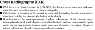
Open itImaging modalties of the Aorta Chest Radiography (CXR) CXR has overall limited sensitivity (~30–60 %) for thoracic aortic aneurysm, and alone cannot be used to exclude acute or chronic aortopathy Calcification or tortuosity of the ascending, arch, and descending thoracic aorta may be visualized, but this is a non-specificific finding in the elderly. Opac
Original toplevel document (pdf)
cannot see any pdfsFlashcard 3628156718348
| status | not learned | measured difficulty | 37% [default] | last interval [days] | |||
|---|---|---|---|---|---|---|---|
| repetition number in this series | 0 | memorised on | scheduled repetition | ||||
| scheduled repetition interval | last repetition or drill |
Parent (intermediate) annotation
Open itaging modalties of the Aorta Chest Radiography (CXR) CXR has overall limited sensitivity (~30–60 %) for thoracic aortic aneurysm, and alone cannot be used to exclude acute or chronic aortopathy <span>Calcification or tortuosity of the ascending, arch, and descending thoracic aorta may be visualized, but this is a non-specificific finding in the elderly. Opacification of the aortico-pulmonary window, enlargement o
Original toplevel document (pdf)
cannot see any pdfsFlashcard 3628158291212
| status | not learned | measured difficulty | 37% [default] | last interval [days] | |||
|---|---|---|---|---|---|---|---|
| repetition number in this series | 0 | memorised on | scheduled repetition | ||||
| scheduled repetition interval | last repetition or drill |
Parent (intermediate) annotation
Open itortic aneurysm, and alone cannot be used to exclude acute or chronic aortopathy Calcification or tortuosity of the ascending, arch, and descending thoracic aorta may be visualized, but this is a <span>non-specificific finding in the elderly. Opacification of the aortico-pulmonary window, enlargement of the thoracic aorta, increased mediastinal width, displacement of trachea from midline, or obscured/i
Original toplevel document (pdf)
cannot see any pdfsFlashcard 3628159864076
| status | not learned | measured difficulty | 37% [default] | last interval [days] | |||
|---|---|---|---|---|---|---|---|
| repetition number in this series | 0 | memorised on | scheduled repetition | ||||
| scheduled repetition interval | last repetition or drill |
Parent (intermediate) annotation
Open itd to exclude acute or chronic aortopathy Calcification or tortuosity of the ascending, arch, and descending thoracic aorta may be visualized, but this is a non-specificific finding in the elderly. <span>Opacification of the aortico-pulmonary window, enlargement of the thoracic aorta, increased mediastinal width, displacement of trachea from midline, or obscured/irregular aortic margin may indicate t
Original toplevel document (pdf)
cannot see any pdfsFlashcard 3628161436940
| status | not learned | measured difficulty | 37% [default] | last interval [days] | |||
|---|---|---|---|---|---|---|---|
| repetition number in this series | 0 | memorised on | scheduled repetition | ||||
| scheduled repetition interval | last repetition or drill |
Parent (intermediate) annotation
Open itfication or tortuosity of the ascending, arch, and descending thoracic aorta may be visualized, but this is a non-specificific finding in the elderly. Opacification of the aortico-pulmonary window, <span>enlargement of the thoracic aorta, increased mediastinal width, displacement of trachea from midline, or obscured/irregular aortic margin may indicate thoracic aortic aneurysm, dissection, or ruptu
Original toplevel document (pdf)
cannot see any pdfsFlashcard 3628163009804
| status | not learned | measured difficulty | 37% [default] | last interval [days] | |||
|---|---|---|---|---|---|---|---|
| repetition number in this series | 0 | memorised on | scheduled repetition | ||||
| scheduled repetition interval | last repetition or drill |
Parent (intermediate) annotation
Open iting, arch, and descending thoracic aorta may be visualized, but this is a non-specificific finding in the elderly. Opacification of the aortico-pulmonary window, enlargement of the thoracic aorta, <span>increased mediastinal width, displacement of trachea from midline, or obscured/irregular aortic margin may indicate thoracic aortic aneurysm, dissection, or rupture. Displaced intimal calcium and
Original toplevel document (pdf)
cannot see any pdfsFlashcard 3628164582668
| status | not learned | measured difficulty | 37% [default] | last interval [days] | |||
|---|---|---|---|---|---|---|---|
| repetition number in this series | 0 | memorised on | scheduled repetition | ||||
| scheduled repetition interval | last repetition or drill |
Parent (intermediate) annotation
Open itracic aorta may be visualized, but this is a non-specificific finding in the elderly. Opacification of the aortico-pulmonary window, enlargement of the thoracic aorta, increased mediastinal width, <span>displacement of trachea from midline, or obscured/irregular aortic margin may indicate thoracic aortic aneurysm, dissection, or rupture. Displaced intimal calcium and pleural effusion may indicate d
Original toplevel document (pdf)
cannot see any pdfsFlashcard 3628166155532
| status | not learned | measured difficulty | 37% [default] | last interval [days] | |||
|---|---|---|---|---|---|---|---|
| repetition number in this series | 0 | memorised on | scheduled repetition | ||||
| scheduled repetition interval | last repetition or drill |
Parent (intermediate) annotation
Open its a non-specificific finding in the elderly. Opacification of the aortico-pulmonary window, enlargement of the thoracic aorta, increased mediastinal width, displacement of trachea from midline, or <span>obscured/irregular aortic margin may indicate thoracic aortic aneurysm, dissection, or rupture. Displaced intimal calcium and pleural effusion may indicate dissection. <span>
Original toplevel document (pdf)
cannot see any pdfsFlashcard 3628167728396
| status | not learned | measured difficulty | 37% [default] | last interval [days] | |||
|---|---|---|---|---|---|---|---|
| repetition number in this series | 0 | memorised on | scheduled repetition | ||||
| scheduled repetition interval | last repetition or drill |
Parent (intermediate) annotation
Open ite thoracic aorta, increased mediastinal width, displacement of trachea from midline, or obscured/irregular aortic margin may indicate thoracic aortic aneurysm, dissection, or rupture. Displaced <span>intimal calcium and pleural effusion may indicate dissection. <span>
Original toplevel document (pdf)
cannot see any pdfs| status | not read | reprioritisations | ||
|---|---|---|---|---|
| last reprioritisation on | suggested re-reading day | |||
| started reading on | finished reading on |
Flashcard 3628175068428
| status | not learned | measured difficulty | 37% [default] | last interval [days] | |||
|---|---|---|---|---|---|---|---|
| repetition number in this series | 0 | memorised on | scheduled repetition | ||||
| scheduled repetition interval | last repetition or drill |
Parent (intermediate) annotation
Open itEchocardiography and Ultrasonography Portable, avoids radiation and contrast media, and can be deployed intra-operatively. Transthoracic Echocardiography (TTE) TTE cannot provide a comprehensive exam of the aorta, but certain regions can be visualized: aortic valve and root, ascending aorta, arch, descending, and abdominal aorta TTE is reasonable for ass
Original toplevel document (pdf)
cannot see any pdfsFlashcard 3628176641292
| status | not learned | measured difficulty | 37% [default] | last interval [days] | |||
|---|---|---|---|---|---|---|---|
| repetition number in this series | 0 | memorised on | scheduled repetition | ||||
| scheduled repetition interval | last repetition or drill |
Parent (intermediate) annotation
Open itt provide a comprehensive exam of the aorta, but certain regions can be visualized: aortic valve and root, ascending aorta, arch, descending, and abdominal aorta TTE is reasonable for assessing <span>aortic valve disorders and monitoring aortic root and ascending aortic dilatation (e.g. especially in Marfan syndrome). It is not sensitive enough to rule out thoracic aortic dissection (sensitivity 70 %). Tr
Original toplevel document (pdf)
cannot see any pdfsFlashcard 3628178214156
| status | not learned | measured difficulty | 37% [default] | last interval [days] | |||
|---|---|---|---|---|---|---|---|
| repetition number in this series | 0 | memorised on | scheduled repetition | ||||
| scheduled repetition interval | last repetition or drill |
Parent (intermediate) annotation
Open itvisualized: aortic valve and root, ascending aorta, arch, descending, and abdominal aorta TTE is reasonable for assessing aortic valve disorders and monitoring aortic root and ascending aortic <span>dilatation (e.g. especially in Marfan syndrome). It is not sensitive enough to rule out thoracic aortic dissection (sensitivity 70 %). Transesophageal Echocardiography (TEE) TEE can visualize the
Original toplevel document (pdf)
cannot see any pdfsFlashcard 3628179787020
| status | not learned | measured difficulty | 37% [default] | last interval [days] | |||
|---|---|---|---|---|---|---|---|
| repetition number in this series | 0 | memorised on | scheduled repetition | ||||
| scheduled repetition interval | last repetition or drill |
Parent (intermediate) annotation
Open itable for assessing aortic valve disorders and monitoring aortic root and ascending aortic dilatation (e.g. especially in Marfan syndrome). It is not sensitive enough to rule out thoracic aortic <span>dissection (sensitivity 70 %). Transesophageal Echocardiography (TEE) TEE can visualize the ascending aorta, transverse arch, and entire descending thoracic aorta. The distal ascending aorta and p
Original toplevel document (pdf)
cannot see any pdfsFlashcard 3628181359884
| status | not learned | measured difficulty | 37% [default] | last interval [days] | |||
|---|---|---|---|---|---|---|---|
| repetition number in this series | 0 | memorised on | scheduled repetition | ||||
| scheduled repetition interval | last repetition or drill |
Parent (intermediate) annotation
Open itc valve disorders and monitoring aortic root and ascending aortic dilatation (e.g. especially in Marfan syndrome). It is not sensitive enough to rule out thoracic aortic dissection (sensitivity <span>70 %). Transesophageal Echocardiography (TEE) TEE can visualize the ascending aorta, transverse arch, and entire descending thoracic aorta. The distal ascending aorta and proximal aortic a
Original toplevel document (pdf)
cannot see any pdfsFlashcard 3628182932748
| status | not learned | measured difficulty | 37% [default] | last interval [days] | |||
|---|---|---|---|---|---|---|---|
| repetition number in this series | 0 | memorised on | scheduled repetition | ||||
| scheduled repetition interval | last repetition or drill |
Parent (intermediate) annotation
Open itndrome). It is not sensitive enough to rule out thoracic aortic dissection (sensitivity 70 %). Transesophageal Echocardiography (TEE) TEE can visualize the ascending aorta, transverse arch, and <span>entire descending thoracic aorta. The distal ascending aorta and proximal aortic arch may be obscured by the trachea. TEE, in contrast to other modalities, can provide functional information such as flow–
Original toplevel document (pdf)
cannot see any pdfsFlashcard 3628184505612
| status | not learned | measured difficulty | 37% [default] | last interval [days] | |||
|---|---|---|---|---|---|---|---|
| repetition number in this series | 0 | memorised on | scheduled repetition | ||||
| scheduled repetition interval | last repetition or drill |
Parent (intermediate) annotation
Open itl Echocardiography (TEE) TEE can visualize the ascending aorta, transverse arch, and entire descending thoracic aorta. The distal ascending aorta and proximal aortic arch may be obscured by the <span>trachea. TEE, in contrast to other modalities, can provide functional information such as flow–dynamics in true and false lumens, detection of AI, detection of cardiac tamponade, and assessment
Original toplevel document (pdf)
cannot see any pdfsFlashcard 3628186078476
| status | not learned | measured difficulty | 37% [default] | last interval [days] | |||
|---|---|---|---|---|---|---|---|
| repetition number in this series | 0 | memorised on | scheduled repetition | ||||
| scheduled repetition interval | last repetition or drill |
Parent (intermediate) annotation
Open ite descending thoracic aorta. The distal ascending aorta and proximal aortic arch may be obscured by the trachea. TEE, in contrast to other modalities, can provide functional information such as <span>flow–dynamics in true and false lumens, detection of AI, detection of cardiac tamponade, and assessment of left ventricular function. Abdominal ultrasonography the technique of choice for screening f
Original toplevel document (pdf)
cannot see any pdfsFlashcard 3628188437772
| status | not learned | measured difficulty | 37% [default] | last interval [days] | |||
|---|---|---|---|---|---|---|---|
| repetition number in this series | 0 | memorised on | scheduled repetition | ||||
| scheduled repetition interval | last repetition or drill |
Parent (intermediate) annotation
Open itrta and proximal aortic arch may be obscured by the trachea. TEE, in contrast to other modalities, can provide functional information such as flow–dynamics in true and false lumens, detection of <span>IAo, detection of cardiac tamponade, and assessment of left ventricular function. Abdominal ultrasonography the technique of choice for screening for infrarenal abdominal aortic aneurysm (A
Original toplevel document (pdf)
cannot see any pdfsFlashcard 3628190010636
| status | not learned | measured difficulty | 37% [default] | last interval [days] | |||
|---|---|---|---|---|---|---|---|
| repetition number in this series | 0 | memorised on | scheduled repetition | ||||
| scheduled repetition interval | last repetition or drill |
Parent (intermediate) annotation
Open itortic arch may be obscured by the trachea. TEE, in contrast to other modalities, can provide functional information such as flow–dynamics in true and false lumens, detection of IAo, detection of <span>cardiac tamponade, and assessment of left ventricular function. Abdominal ultrasonography the technique of choice for screening for infrarenal abdominal aortic aneurysm (AAA), but is less accurate as app
Original toplevel document (pdf)
cannot see any pdfsFlashcard 3628191583500
| status | not learned | measured difficulty | 37% [default] | last interval [days] | |||
|---|---|---|---|---|---|---|---|
| repetition number in this series | 0 | memorised on | scheduled repetition | ||||
| scheduled repetition interval | last repetition or drill |
Parent (intermediate) annotation
Open itchea. TEE, in contrast to other modalities, can provide functional information such as flow–dynamics in true and false lumens, detection of IAo, detection of cardiac tamponade, and assessment of <span>left ventricular function. Abdominal ultrasonography the technique of choice for screening for infrarenal abdominal aortic aneurysm (AAA), but is less accurate as applied to the suprarenal aorta or branch vessel
Original toplevel document (pdf)
cannot see any pdfsFlashcard 3628193156364
| status | not learned | measured difficulty | 37% [default] | last interval [days] | |||
|---|---|---|---|---|---|---|---|
| repetition number in this series | 0 | memorised on | scheduled repetition | ||||
| scheduled repetition interval | last repetition or drill |
Parent (intermediate) annotation
Open itmics in true and false lumens, detection of IAo, detection of cardiac tamponade, and assessment of left ventricular function. Abdominal ultrasonography the technique of choice for screening for <span>infrarenal abdominal aortic aneurysm (AAA), but is less accurate as applied to the suprarenal aorta or branch vessels. <span>
Original toplevel document (pdf)
cannot see any pdfsFlashcard 3628194729228
| status | not learned | measured difficulty | 37% [default] | last interval [days] | |||
|---|---|---|---|---|---|---|---|
| repetition number in this series | 0 | memorised on | scheduled repetition | ||||
| scheduled repetition interval | last repetition or drill |
Parent (intermediate) annotation
Open itd assessment of left ventricular function. Abdominal ultrasonography the technique of choice for screening for infrarenal abdominal aortic aneurysm (AAA), but is less accurate as applied to the <span>suprarenal aorta or branch vessels. <span>
Original toplevel document (pdf)
cannot see any pdfs| status | not read | reprioritisations | ||
|---|---|---|---|---|
| last reprioritisation on | suggested re-reading day | |||
| started reading on | finished reading on |
Flashcard 3628214914316
| status | not learned | measured difficulty | 37% [default] | last interval [days] | |||
|---|---|---|---|---|---|---|---|
| repetition number in this series | 0 | memorised on | scheduled repetition | ||||
| scheduled repetition interval | last repetition or drill |
Parent (intermediate) annotation
Open itImaging modalties of the Aorta Computed Tomography (CT) CT is a highly accurate, rapid, reproducible, and readily available technique for detecting and sizing aortic aneurysms and for the diagnostic evaluation of suspected aortic dissection. CT is also helpful at m
Original toplevel document (pdf)
cannot see any pdfsFlashcard 3628216487180
| status | not learned | measured difficulty | 37% [default] | last interval [days] | |||
|---|---|---|---|---|---|---|---|
| repetition number in this series | 0 | memorised on | scheduled repetition | ||||
| scheduled repetition interval | last repetition or drill |
Parent (intermediate) annotation
Open itImaging modalties of the Aorta Computed Tomography (CT) CT is a highly accurate, rapid, reproducible, and readily available technique for detecting and sizing aortic aneurysms and for the diagnostic evaluation of suspected aortic dissection. CT is also helpful at mapping branch vessels, and for detecting mimics of aortic disease (e
Original toplevel document (pdf)
cannot see any pdfsFlashcard 3628218060044
| status | not learned | measured difficulty | 37% [default] | last interval [days] | |||
|---|---|---|---|---|---|---|---|
| repetition number in this series | 0 | memorised on | scheduled repetition | ||||
| scheduled repetition interval | last repetition or drill |
Parent (intermediate) annotation
Open itspan> Imaging modalties of the Aorta Computed Tomography (CT) CT is a highly accurate, rapid, reproducible, and readily available technique for detecting and sizing aortic aneurysms and for the <span>diagnostic evaluation of suspected aortic dissection. CT is also helpful at mapping branch vessels, and for detecting mimics of aortic disease (e.g. pericardial disease, gastrointestinal disease).
Original toplevel document (pdf)
cannot see any pdfsFlashcard 3628219632908
| status | not learned | measured difficulty | 37% [default] | last interval [days] | |||
|---|---|---|---|---|---|---|---|
| repetition number in this series | 0 | memorised on | scheduled repetition | ||||
| scheduled repetition interval | last repetition or drill |
Parent (intermediate) annotation
Open itImaging modalties of the Aorta Computed Tomography (CT) CT is a highly accurate, rapid, reproducible, and readily available technique for detecting and sizing aortic aneurysms and for the diagnostic evaluation of suspected aortic dissection. CT is also helpful at mapping branch vessels, and for detecting mimics of aortic disease (e.g. pericar
Original toplevel document (pdf)
cannot see any pdfsFlashcard 3628221205772
| status | not learned | measured difficulty | 37% [default] | last interval [days] | |||
|---|---|---|---|---|---|---|---|
| repetition number in this series | 0 | memorised on | scheduled repetition | ||||
| scheduled repetition interval | last repetition or drill |
Parent (intermediate) annotation
Open itaccurate, rapid, reproducible, and readily available technique for detecting and sizing aortic aneurysms and for the diagnostic evaluation of suspected aortic dissection. CT is also helpful at <span>mapping branch vessels, and for detecting mimics of aortic disease (e.g. pericardial disease, gastrointestinal disease). Magnetic Resonance (MR) Imaging MR is also a highly accurate technique f
Original toplevel document (pdf)
cannot see any pdfsFlashcard 3628222778636
| status | not learned | measured difficulty | 37% [default] | last interval [days] | |||
|---|---|---|---|---|---|---|---|
| repetition number in this series | 0 | memorised on | scheduled repetition | ||||
| scheduled repetition interval | last repetition or drill |
Parent (intermediate) annotation
Open itting and sizing aortic aneurysms and for the diagnostic evaluation of suspected aortic dissection. CT is also helpful at mapping branch vessels, and for detecting mimics of aortic disease (e.g. <span>pericardial disease, gastrointestinal disease). Magnetic Resonance (MR) Imaging MR is also a highly accurate technique for aortic imaging. However, the study time is lengthy and the patient is relatively i
Original toplevel document (pdf)
cannot see any pdfsFlashcard 3628224351500
| status | not learned | measured difficulty | 37% [default] | last interval [days] | |||
|---|---|---|---|---|---|---|---|
| repetition number in this series | 0 | memorised on | scheduled repetition | ||||
| scheduled repetition interval | last repetition or drill |
Parent (intermediate) annotation
Open itc aneurysms and for the diagnostic evaluation of suspected aortic dissection. CT is also helpful at mapping branch vessels, and for detecting mimics of aortic disease (e.g. pericardial disease, <span>gastrointestinal disease). Magnetic Resonance (MR) Imaging MR is also a highly accurate technique for aortic imaging. However, the study time is lengthy and the patient is relatively inaccessible, making this m
Original toplevel document (pdf)
cannot see any pdfsFlashcard 3628225924364
| status | not learned | measured difficulty | 37% [default] | last interval [days] | |||
|---|---|---|---|---|---|---|---|
| repetition number in this series | 0 | memorised on | scheduled repetition | ||||
| scheduled repetition interval | last repetition or drill |
Parent (intermediate) annotation
Open itT is also helpful at mapping branch vessels, and for detecting mimics of aortic disease (e.g. pericardial disease, gastrointestinal disease). Magnetic Resonance (MR) Imaging MR is also a highly <span>accurate technique for aortic imaging. However, the study time is lengthy and the patient is relatively inaccessible, making this modality unsuitable for acute or unstable patients. MR is most o
Original toplevel document (pdf)
cannot see any pdfsFlashcard 3628227497228
| status | not learned | measured difficulty | 37% [default] | last interval [days] | |||
|---|---|---|---|---|---|---|---|
| repetition number in this series | 0 | memorised on | scheduled repetition | ||||
| scheduled repetition interval | last repetition or drill |
Parent (intermediate) annotation
Open it). Magnetic Resonance (MR) Imaging MR is also a highly accurate technique for aortic imaging. However, the study time is lengthy and the patient is relatively inaccessible, making this modality <span>unsuitable for acute or unstable patients. MR is most often performed with intravenous gadolinium as a contrast agent, but the “black-blood” technique with spin-echo sequences can provide satisfac
Original toplevel document (pdf)
cannot see any pdfsFlashcard 3628229070092
| status | not learned | measured difficulty | 37% [default] | last interval [days] | |||
|---|---|---|---|---|---|---|---|
| repetition number in this series | 0 | memorised on | scheduled repetition | ||||
| scheduled repetition interval | last repetition or drill |
Parent (intermediate) annotation
Open itaging. However, the study time is lengthy and the patient is relatively inaccessible, making this modality unsuitable for acute or unstable patients. MR is most often performed with intravenous <span>gadolinium as a contrast agent, but the “black-blood” technique with spin-echo sequences can provide satisfactory images without the need for gadolinium. Aortography Catheter-based aortography is
Original toplevel document (pdf)
cannot see any pdfsFlashcard 3628230642956
| status | not learned | measured difficulty | 37% [default] | last interval [days] | |||
|---|---|---|---|---|---|---|---|
| repetition number in this series | 0 | memorised on | scheduled repetition | ||||
| scheduled repetition interval | last repetition or drill |
Parent (intermediate) annotation
Open itood” technique with spin-echo sequences can provide satisfactory images without the need for gadolinium. Aortography Catheter-based aortography is an invasive technique that can demonstrate the <span>full extent of aneurysmal disease and dissection, map branch vessel involvement, and demonstrate the presence of IAo. However, aortography is not readily available in most settings, requires an exp