Edited, memorised or added to reading queue
on 20-Nov-2016 (Sun)
Do you want BuboFlash to help you learning these things? Click here to log in or create user.
| status | not read | reprioritisations | ||
|---|---|---|---|---|
| last reprioritisation on | suggested re-reading day | |||
| started reading on | finished reading on |
| status | not read | reprioritisations | ||
|---|---|---|---|---|
| last reprioritisation on | suggested re-reading day | |||
| started reading on | finished reading on |
Subject 3. Market Equilibrium
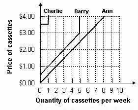
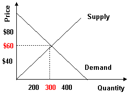
Aggregate Demand and Aggregate Supply An aggregate demand curve is simply a schedule that shows amounts of a product that buyers collectively desire to purchase at each possible price level. An aggregate supply curve is simply a curve showing the amounts of a product that all firms will produce at each price level. Example 1 Refer to the graph below. What is the market quantity that would be supplied at a price of $2.00? Market quantity is the sum of individual quantities supplied at each price. At a price of $2.00, Ann supplies 4, Barry supplies 3, and Charlie supplies 0. The market supply is 7. Market Equilibrium Equilibrium is a state in which conflicting forces are in balance. In equilibrium, it will be possible for both buyers and sel
| status | not read | reprioritisations | ||
|---|---|---|---|---|
| last reprioritisation on | suggested re-reading day | |||
| started reading on | finished reading on |
Subject 3. Market Equilibrium
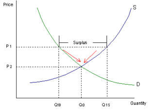
; Market quantity is the sum of individual quantities supplied at each price. At a price of $2.00, Ann supplies 4, Barry supplies 3, and Charlie supplies 0. The market supply is 7. <span>Market Equilibrium Equilibrium is a state in which conflicting forces are in balance. In equilibrium, it will be possible for both buyers and sellers to realize their goals simultaneously. The following graph depicts the market supply and demand for concert tickets at Madison Square Garden in New York City. Equilibrium price and quantity are where the supply and demand curves intersect. Draw a horizontal line from the intersection to the price axis. This is equilibrium price: $60. Draw a vertical line from the intersection to the quantity axis. This is equilibrium quantity: 300. It is equilibrium because quantity demanded equals quantity supplied at $60 per ticket. At this price, there is neither surplus (excess supply) nor shortage (excess demand), so there is no downward or upward pressure for the price to change. Surplus will push prices downward towards equilibrium. Suppose the price is initially above the equilibrium price (P 2 ) and sits at P 1 . Quantity supplied (Q 1s ) will exceed quantity demanded (Q 1D ), creating a surplus. The surplus will put downward pressure on prices since producers will begin to lower their prices to sell the surplus. As a result, the price will fall, the quantity supplied will decrease, and the quantity demanded will increase until the equilibrium price (P 2 ) is restored. This process involves movements along supply-and-demand curves since the changes are caused by price fluctuations. Similarly, shortages push prices upward towards equilibrium. Because the price rises if it is below equilibrium, falls if it is above equilibrium, and remains constant if it is at equilibrium, the price is pulled toward equilibrium and remains there until some event changes the equilibrium. We refer to such an equilibrium as being stable because whenever price is disturbed away from the equilibrium, it tends to converge back to that equilibrium. An unstable equilibrium is an equilibrium that is not restored if disrupted by an external force. While most equilibria studied in economics are of the stable variety, a few cases of unstable equilibria do emerge from time to time, in limited circumstances.<span><body><html>
| status | not read | reprioritisations | ||
|---|---|---|---|---|
| last reprioritisation on | suggested re-reading day | |||
| started reading on | finished reading on |
Parent (intermediate) annotation
Open itM2-receptors modulate muscarinic potassium channels. [11] [12] In the heart, this contributes to a decreased heart rate. They do so by the G beta gamma subunit of the G protein coupled to M 2 . This part of the G protein can open K + channels in the parasympathetic notches in the heart, which causes an outward current of potassium, which slows down the heart rate.
Original toplevel document
Muscarinic acetylcholine receptor M2 - Wikipediais claim instead found no significant association between the CHRM2 gene and intelligence. [8] Olfactory behavior[edit] Mediating olfactory guided behaviors (e.g. odor discrimination, aggression, mating) [9] Mechanism of action[edit] <span>M 2 muscarinic receptors act via a G i type receptor, which causes a decrease in cAMP in the cell, generally leading to inhibitory-type effects. They appear to serve as autoreceptors. [10] In addition, they modulate muscarinic potassium channels. [11] [12] In the heart, this contributes to a decreased heart rate. They do so by the G beta gamma subunit of the G protein coupled to M 2 . This part of the G protein can open K + channels in the parasympathetic notches in the heart, which causes an outward current of potassium, which slows down the heart rate. Ligands[edit] Few highly selective M 2 agonists are available at present, although there are several non-selective muscarinic agonists that stimulate M 2 , and a number of selectiv
| status | not read | reprioritisations | ||
|---|---|---|---|---|
| last reprioritisation on | suggested re-reading day | |||
| started reading on | finished reading on |
Flashcard 1420469079308
| status | not learned | measured difficulty | 37% [default] | last interval [days] | |||
|---|---|---|---|---|---|---|---|
| repetition number in this series | 0 | memorised on | scheduled repetition | ||||
| scheduled repetition interval | last repetition or drill |
Parent (intermediate) annotation
Open itPompy ciepła wykorzystują zjawisko przepływu ciepła.
Original toplevel document (pdf)
cannot see any pdfsFlashcard 1420470652172
| status | not learned | measured difficulty | 37% [default] | last interval [days] | |||
|---|---|---|---|---|---|---|---|
| repetition number in this series | 0 | memorised on | scheduled repetition | ||||
| scheduled repetition interval | last repetition or drill |
Parent (intermediate) annotation
Open itPompy ciepła wykorzystują zjawisko przepływu ciepła.
Original toplevel document (pdf)
cannot see any pdfs| status | not read | reprioritisations | ||
|---|---|---|---|---|
| last reprioritisation on | suggested re-reading day | |||
| started reading on | finished reading on |
Parent (intermediate) annotation
Open itPompy ciepła wykorzystują zjawisko przepływu ciepła. Pompy ciepła wykorzystują zjawisko zmiany stanu fizycznego substancji. Przykład. Jeżeli do gotującej się w naczyniu wody będziemy dalej dostarczać energię (ciepło), to cała woda wyparuje, a jej temperatura nie wzrośnie. Ilość energii związana ze
Original toplevel document (pdf)
cannot see any pdfs| status | not read | reprioritisations | ||
|---|---|---|---|---|
| last reprioritisation on | suggested re-reading day | |||
| started reading on | finished reading on |
| status | not read | reprioritisations | ||
|---|---|---|---|---|
| last reprioritisation on | suggested re-reading day | |||
| started reading on | finished reading on |
| status | not read | reprioritisations | ||
|---|---|---|---|---|
| last reprioritisation on | suggested re-reading day | |||
| started reading on | finished reading on |
Parent (intermediate) annotation
Open it/head>Entalpia – określa zawartość energii w układzie termodynamicznym, oznaczana jako: H, I [kJ; Wh]; entalpia właściwa: h, i [kJ/kg, Wh/kg]. Entalpia, ze starogreckiego: „en” – „w” i „thalpein” – „rozgrzewać”. entalpia to zawartość energii <html>
Original toplevel document (pdf)
cannot see any pdfs| status | not read | reprioritisations | ||
|---|---|---|---|---|
| last reprioritisation on | suggested re-reading day | |||
| started reading on | finished reading on |
| status | not read | reprioritisations | ||
|---|---|---|---|---|
| last reprioritisation on | suggested re-reading day | |||
| started reading on | finished reading on |
| status | not read | reprioritisations | ||
|---|---|---|---|---|
| last reprioritisation on | suggested re-reading day | |||
| started reading on | finished reading on |
| status | not read | reprioritisations | ||
|---|---|---|---|---|
| last reprioritisation on | suggested re-reading day | |||
| started reading on | finished reading on |
| status | not read | reprioritisations | ||
|---|---|---|---|---|
| last reprioritisation on | suggested re-reading day | |||
| started reading on | finished reading on |
Flashcard 1420723621132
| status | not learned | measured difficulty | 37% [default] | last interval [days] | |||
|---|---|---|---|---|---|---|---|
| repetition number in this series | 0 | memorised on | scheduled repetition | ||||
| scheduled repetition interval | last repetition or drill |
Parent (intermediate) annotation
Open itM2-receptorer decreases heartrate by the G beta gamma subunit of the G protein coupled to M 2 . This part of the G protein can open K + channels, which causes an outward current of potassium, which slows down the heart rate.
Original toplevel document
Muscarinic acetylcholine receptor M2 - Wikipediais claim instead found no significant association between the CHRM2 gene and intelligence. [8] Olfactory behavior[edit] Mediating olfactory guided behaviors (e.g. odor discrimination, aggression, mating) [9] Mechanism of action[edit] <span>M 2 muscarinic receptors act via a G i type receptor, which causes a decrease in cAMP in the cell, generally leading to inhibitory-type effects. They appear to serve as autoreceptors. [10] In addition, they modulate muscarinic potassium channels. [11] [12] In the heart, this contributes to a decreased heart rate. They do so by the G beta gamma subunit of the G protein coupled to M 2 . This part of the G protein can open K + channels in the parasympathetic notches in the heart, which causes an outward current of potassium, which slows down the heart rate. Ligands[edit] Few highly selective M 2 agonists are available at present, although there are several non-selective muscarinic agonists that stimulate M 2 , and a number of selectiv
| status | not read | reprioritisations | ||
|---|---|---|---|---|
| last reprioritisation on | suggested re-reading day | |||
| started reading on | finished reading on |
Parent (intermediate) annotation
Open itM2-receptorer decreases heartrate by the G beta gamma subunit of the G protein coupled to M 2 . This part of the G protein can open K + channels, which causes an outward current of potassium, which slows down the heart rate.
Original toplevel document
Muscarinic acetylcholine receptor M2 - Wikipediais claim instead found no significant association between the CHRM2 gene and intelligence. [8] Olfactory behavior[edit] Mediating olfactory guided behaviors (e.g. odor discrimination, aggression, mating) [9] Mechanism of action[edit] <span>M 2 muscarinic receptors act via a G i type receptor, which causes a decrease in cAMP in the cell, generally leading to inhibitory-type effects. They appear to serve as autoreceptors. [10] In addition, they modulate muscarinic potassium channels. [11] [12] In the heart, this contributes to a decreased heart rate. They do so by the G beta gamma subunit of the G protein coupled to M 2 . This part of the G protein can open K + channels in the parasympathetic notches in the heart, which causes an outward current of potassium, which slows down the heart rate. Ligands[edit] Few highly selective M 2 agonists are available at present, although there are several non-selective muscarinic agonists that stimulate M 2 , and a number of selectiv
Flashcard 1420728601868
| status | not learned | measured difficulty | 37% [default] | last interval [days] | |||
|---|---|---|---|---|---|---|---|
| repetition number in this series | 0 | memorised on | scheduled repetition | ||||
| scheduled repetition interval | last repetition or drill |
Parent (intermediate) annotation
Open itM2-receptorer decreases heartrate by the G beta gamma subunit of the G protein coupled to M 2 .
Original toplevel document
Muscarinic acetylcholine receptor M2 - Wikipediais claim instead found no significant association between the CHRM2 gene and intelligence. [8] Olfactory behavior[edit] Mediating olfactory guided behaviors (e.g. odor discrimination, aggression, mating) [9] Mechanism of action[edit] <span>M 2 muscarinic receptors act via a G i type receptor, which causes a decrease in cAMP in the cell, generally leading to inhibitory-type effects. They appear to serve as autoreceptors. [10] In addition, they modulate muscarinic potassium channels. [11] [12] In the heart, this contributes to a decreased heart rate. They do so by the G beta gamma subunit of the G protein coupled to M 2 . This part of the G protein can open K + channels in the parasympathetic notches in the heart, which causes an outward current of potassium, which slows down the heart rate. Ligands[edit] Few highly selective M 2 agonists are available at present, although there are several non-selective muscarinic agonists that stimulate M 2 , and a number of selectiv
Flashcard 1420730174732
| status | not learned | measured difficulty | 37% [default] | last interval [days] | |||
|---|---|---|---|---|---|---|---|
| repetition number in this series | 0 | memorised on | scheduled repetition | ||||
| scheduled repetition interval | last repetition or drill |
Parent (intermediate) annotation
Open itM2-receptorer decreases heartrate by the G beta gamma subunit of the G protein coupled to M 2 .
Original toplevel document
Muscarinic acetylcholine receptor M2 - Wikipediais claim instead found no significant association between the CHRM2 gene and intelligence. [8] Olfactory behavior[edit] Mediating olfactory guided behaviors (e.g. odor discrimination, aggression, mating) [9] Mechanism of action[edit] <span>M 2 muscarinic receptors act via a G i type receptor, which causes a decrease in cAMP in the cell, generally leading to inhibitory-type effects. They appear to serve as autoreceptors. [10] In addition, they modulate muscarinic potassium channels. [11] [12] In the heart, this contributes to a decreased heart rate. They do so by the G beta gamma subunit of the G protein coupled to M 2 . This part of the G protein can open K + channels in the parasympathetic notches in the heart, which causes an outward current of potassium, which slows down the heart rate. Ligands[edit] Few highly selective M 2 agonists are available at present, although there are several non-selective muscarinic agonists that stimulate M 2 , and a number of selectiv
Flashcard 1420731747596
| status | not learned | measured difficulty | 37% [default] | last interval [days] | |||
|---|---|---|---|---|---|---|---|
| repetition number in this series | 0 | memorised on | scheduled repetition | ||||
| scheduled repetition interval | last repetition or drill |
Parent (intermediate) annotation
Open itM2-receptorer decreases heartrate by the G beta gamma subunit of the G protein coupled to M 2 .
Original toplevel document
Muscarinic acetylcholine receptor M2 - Wikipediais claim instead found no significant association between the CHRM2 gene and intelligence. [8] Olfactory behavior[edit] Mediating olfactory guided behaviors (e.g. odor discrimination, aggression, mating) [9] Mechanism of action[edit] <span>M 2 muscarinic receptors act via a G i type receptor, which causes a decrease in cAMP in the cell, generally leading to inhibitory-type effects. They appear to serve as autoreceptors. [10] In addition, they modulate muscarinic potassium channels. [11] [12] In the heart, this contributes to a decreased heart rate. They do so by the G beta gamma subunit of the G protein coupled to M 2 . This part of the G protein can open K + channels in the parasympathetic notches in the heart, which causes an outward current of potassium, which slows down the heart rate. Ligands[edit] Few highly selective M 2 agonists are available at present, although there are several non-selective muscarinic agonists that stimulate M 2 , and a number of selectiv
Flashcard 1420733320460
| status | not learned | measured difficulty | 37% [default] | last interval [days] | |||
|---|---|---|---|---|---|---|---|
| repetition number in this series | 0 | memorised on | scheduled repetition | ||||
| scheduled repetition interval | last repetition or drill |
Parent (intermediate) annotation
Open itAntidiuretic effect of vasopressin occurs via activation of V 2 R.
Original toplevel document
Vasopressin receptor - Wikipedialuminal membrane of the collecting duct limit the antidiuretic action of vasopressin. Additionally, vasopressin selectively contracts efferent arterioles probably through the V 1 R, but not the afferent arteriole. [1] V 2 receptor[edit] <span>V 2 receptor (V 2 R) differs from V 1 R primarily in the number of sites susceptible to N-linked glycosylation; the V 1 R has sites at both the amino-terminus and at the extracellular loop, whereas the V 2 R has a single site at the extracellular amino-terminus. [1] The well known antidiuretic effect of vasopressin occurs via activation of V 2 R. [1] Vasopressin regulates water excretion from the kidney by increasing the osmotic water permeability of the renal collecting duct – an effect that is explained by coupling of the V 2 R with the G s signaling pathway, which activates cAMP. Interestingly, the V 2 R continues to activate G s after being internalized by β-arrestin rather than being desensitized. This internalized G s signaling by V 2 R is explained by the receptors ability to form "mega-complexes" consisting of a single V 2 R, β-arrestin, and heterotrimeric G s . [3] The increased intracellular cAMP in the kidney in turn triggers fusion of aquaporin-2-bearing vesicles with the apical plasma membrane of the collecting duct principal cells, increasing water reabsorption. [1] V 3 receptor[edit] The human V 3 receptor (V 3 R, previously known as V 1B R) is a G-protein-coupled pituitary receptor that, because of its scarcity, was only recently characteri
Flashcard 1420734893324
| status | not learned | measured difficulty | 37% [default] | last interval [days] | |||
|---|---|---|---|---|---|---|---|
| repetition number in this series | 0 | memorised on | scheduled repetition | ||||
| scheduled repetition interval | last repetition or drill |
Parent (intermediate) annotation
Open itAntidiuretic effect of vasopressin occurs via activation of V 2 R.
Original toplevel document
Vasopressin receptor - Wikipedialuminal membrane of the collecting duct limit the antidiuretic action of vasopressin. Additionally, vasopressin selectively contracts efferent arterioles probably through the V 1 R, but not the afferent arteriole. [1] V 2 receptor[edit] <span>V 2 receptor (V 2 R) differs from V 1 R primarily in the number of sites susceptible to N-linked glycosylation; the V 1 R has sites at both the amino-terminus and at the extracellular loop, whereas the V 2 R has a single site at the extracellular amino-terminus. [1] The well known antidiuretic effect of vasopressin occurs via activation of V 2 R. [1] Vasopressin regulates water excretion from the kidney by increasing the osmotic water permeability of the renal collecting duct – an effect that is explained by coupling of the V 2 R with the G s signaling pathway, which activates cAMP. Interestingly, the V 2 R continues to activate G s after being internalized by β-arrestin rather than being desensitized. This internalized G s signaling by V 2 R is explained by the receptors ability to form "mega-complexes" consisting of a single V 2 R, β-arrestin, and heterotrimeric G s . [3] The increased intracellular cAMP in the kidney in turn triggers fusion of aquaporin-2-bearing vesicles with the apical plasma membrane of the collecting duct principal cells, increasing water reabsorption. [1] V 3 receptor[edit] The human V 3 receptor (V 3 R, previously known as V 1B R) is a G-protein-coupled pituitary receptor that, because of its scarcity, was only recently characteri
Flashcard 1420736466188
| status | not learned | measured difficulty | 37% [default] | last interval [days] | |||
|---|---|---|---|---|---|---|---|
| repetition number in this series | 0 | memorised on | scheduled repetition | ||||
| scheduled repetition interval | last repetition or drill |
Parent (intermediate) annotation
Open itAntidiuretic effect of vasopressin occurs via activation of V 2 R.
Original toplevel document
Vasopressin receptor - Wikipedialuminal membrane of the collecting duct limit the antidiuretic action of vasopressin. Additionally, vasopressin selectively contracts efferent arterioles probably through the V 1 R, but not the afferent arteriole. [1] V 2 receptor[edit] <span>V 2 receptor (V 2 R) differs from V 1 R primarily in the number of sites susceptible to N-linked glycosylation; the V 1 R has sites at both the amino-terminus and at the extracellular loop, whereas the V 2 R has a single site at the extracellular amino-terminus. [1] The well known antidiuretic effect of vasopressin occurs via activation of V 2 R. [1] Vasopressin regulates water excretion from the kidney by increasing the osmotic water permeability of the renal collecting duct – an effect that is explained by coupling of the V 2 R with the G s signaling pathway, which activates cAMP. Interestingly, the V 2 R continues to activate G s after being internalized by β-arrestin rather than being desensitized. This internalized G s signaling by V 2 R is explained by the receptors ability to form "mega-complexes" consisting of a single V 2 R, β-arrestin, and heterotrimeric G s . [3] The increased intracellular cAMP in the kidney in turn triggers fusion of aquaporin-2-bearing vesicles with the apical plasma membrane of the collecting duct principal cells, increasing water reabsorption. [1] V 3 receptor[edit] The human V 3 receptor (V 3 R, previously known as V 1B R) is a G-protein-coupled pituitary receptor that, because of its scarcity, was only recently characteri
Flashcard 1420738039052
| status | not learned | measured difficulty | 37% [default] | last interval [days] | |||
|---|---|---|---|---|---|---|---|
| repetition number in this series | 0 | memorised on | scheduled repetition | ||||
| scheduled repetition interval | last repetition or drill |
Parent (intermediate) annotation
Open itAntidiuretic effect of vasopressin occurs via activation of V 2 R.
Original toplevel document
Vasopressin receptor - Wikipedialuminal membrane of the collecting duct limit the antidiuretic action of vasopressin. Additionally, vasopressin selectively contracts efferent arterioles probably through the V 1 R, but not the afferent arteriole. [1] V 2 receptor[edit] <span>V 2 receptor (V 2 R) differs from V 1 R primarily in the number of sites susceptible to N-linked glycosylation; the V 1 R has sites at both the amino-terminus and at the extracellular loop, whereas the V 2 R has a single site at the extracellular amino-terminus. [1] The well known antidiuretic effect of vasopressin occurs via activation of V 2 R. [1] Vasopressin regulates water excretion from the kidney by increasing the osmotic water permeability of the renal collecting duct – an effect that is explained by coupling of the V 2 R with the G s signaling pathway, which activates cAMP. Interestingly, the V 2 R continues to activate G s after being internalized by β-arrestin rather than being desensitized. This internalized G s signaling by V 2 R is explained by the receptors ability to form "mega-complexes" consisting of a single V 2 R, β-arrestin, and heterotrimeric G s . [3] The increased intracellular cAMP in the kidney in turn triggers fusion of aquaporin-2-bearing vesicles with the apical plasma membrane of the collecting duct principal cells, increasing water reabsorption. [1] V 3 receptor[edit] The human V 3 receptor (V 3 R, previously known as V 1B R) is a G-protein-coupled pituitary receptor that, because of its scarcity, was only recently characteri
Flashcard 1420740398348
| status | not learned | measured difficulty | 37% [default] | last interval [days] | |||
|---|---|---|---|---|---|---|---|
| repetition number in this series | 0 | memorised on | scheduled repetition | ||||
| scheduled repetition interval | last repetition or drill |
Parent (intermediate) annotation
Open itV 1 receptors (V 1 Rs) are found in high density on vascular smooth muscle and cause vasoconstriction by an increase in intracellular calcium via the phosphatidyl–inositol-bisphosphonate cascade.
Original toplevel document
Vasopressin receptor - Wikipediaf collecting duct, vascular endothelium and vascular smooth muscle cell insertion of AQP-2 water channels into apical membrane, induction of AQP-2 synthesis, releases von Willebrand factor and factor VIII, vasodilation V 1 receptor[edit] <span>V 1 receptors (V 1 Rs) are found in high density on vascular smooth muscle and cause vasoconstriction by an increase in intracellular calcium via the phosphatidyl–inositol-bisphosphonate cascade. [1] Cardiac myocytes also possess V 1 R. Additionally V 1 R are located in brain, testis, superior cervical ganglion, liver, blood vessels, and renal medulla. [1] V 1 R is present on platelets, which upon stimulation induces an increase in intracellular calcium, facilitating thrombosis. Studies have indicated that due to polymorphism of platelet V 1 R there is significant heterogeneity in the aggregation response of normal human platelets to vasopressin. [1] V 1 Rs are found in kidney, where they occur in high density on medullary interstitial cells, vasa recta, and epithelial cells of the collecting duct. [1] Vasopressin acts on medullary vasculature through V 1 R to reduce blood flow to inner medulla without affecting blood flow to outer medulla. V 1 Rs on the luminal membrane of the collecting duct limit the antidiuretic action of vasopressin. Additionally, vasopressin selectively contracts efferent arterioles probably through the V 1 R, but not the afferent arteriole. [1] V 2 receptor[edit] V 2 receptor (V 2 R) differs from V 1 R primarily in the number of sites susceptible to N-linked glycosylation; the V 1 R has sites at both the amino-terminus a
Flashcard 1420742757644
| status | not learned | measured difficulty | 37% [default] | last interval [days] | |||
|---|---|---|---|---|---|---|---|
| repetition number in this series | 0 | memorised on | scheduled repetition | ||||
| scheduled repetition interval | last repetition or drill |
Parent (intermediate) annotation
Open itV 1 receptors (V 1 Rs) are found in high density on vascular smooth muscle and cause vasoconstriction by an increase in intracellular calcium via the phosphatidyl–inositol-bisphosphonate cascade.
Original toplevel document
Vasopressin receptor - Wikipediaf collecting duct, vascular endothelium and vascular smooth muscle cell insertion of AQP-2 water channels into apical membrane, induction of AQP-2 synthesis, releases von Willebrand factor and factor VIII, vasodilation V 1 receptor[edit] <span>V 1 receptors (V 1 Rs) are found in high density on vascular smooth muscle and cause vasoconstriction by an increase in intracellular calcium via the phosphatidyl–inositol-bisphosphonate cascade. [1] Cardiac myocytes also possess V 1 R. Additionally V 1 R are located in brain, testis, superior cervical ganglion, liver, blood vessels, and renal medulla. [1] V 1 R is present on platelets, which upon stimulation induces an increase in intracellular calcium, facilitating thrombosis. Studies have indicated that due to polymorphism of platelet V 1 R there is significant heterogeneity in the aggregation response of normal human platelets to vasopressin. [1] V 1 Rs are found in kidney, where they occur in high density on medullary interstitial cells, vasa recta, and epithelial cells of the collecting duct. [1] Vasopressin acts on medullary vasculature through V 1 R to reduce blood flow to inner medulla without affecting blood flow to outer medulla. V 1 Rs on the luminal membrane of the collecting duct limit the antidiuretic action of vasopressin. Additionally, vasopressin selectively contracts efferent arterioles probably through the V 1 R, but not the afferent arteriole. [1] V 2 receptor[edit] V 2 receptor (V 2 R) differs from V 1 R primarily in the number of sites susceptible to N-linked glycosylation; the V 1 R has sites at both the amino-terminus a
Flashcard 1420744330508
| status | not learned | measured difficulty | 37% [default] | last interval [days] | |||
|---|---|---|---|---|---|---|---|
| repetition number in this series | 0 | memorised on | scheduled repetition | ||||
| scheduled repetition interval | last repetition or drill |
Parent (intermediate) annotation
Open itV 1 receptors (V 1 Rs) are found in high density on vascular smooth muscle and cause vasoconstriction by an increase in intracellular calcium via the phosphatidyl–inositol-bisphosphonate cascade.
Original toplevel document
Vasopressin receptor - Wikipediaf collecting duct, vascular endothelium and vascular smooth muscle cell insertion of AQP-2 water channels into apical membrane, induction of AQP-2 synthesis, releases von Willebrand factor and factor VIII, vasodilation V 1 receptor[edit] <span>V 1 receptors (V 1 Rs) are found in high density on vascular smooth muscle and cause vasoconstriction by an increase in intracellular calcium via the phosphatidyl–inositol-bisphosphonate cascade. [1] Cardiac myocytes also possess V 1 R. Additionally V 1 R are located in brain, testis, superior cervical ganglion, liver, blood vessels, and renal medulla. [1] V 1 R is present on platelets, which upon stimulation induces an increase in intracellular calcium, facilitating thrombosis. Studies have indicated that due to polymorphism of platelet V 1 R there is significant heterogeneity in the aggregation response of normal human platelets to vasopressin. [1] V 1 Rs are found in kidney, where they occur in high density on medullary interstitial cells, vasa recta, and epithelial cells of the collecting duct. [1] Vasopressin acts on medullary vasculature through V 1 R to reduce blood flow to inner medulla without affecting blood flow to outer medulla. V 1 Rs on the luminal membrane of the collecting duct limit the antidiuretic action of vasopressin. Additionally, vasopressin selectively contracts efferent arterioles probably through the V 1 R, but not the afferent arteriole. [1] V 2 receptor[edit] V 2 receptor (V 2 R) differs from V 1 R primarily in the number of sites susceptible to N-linked glycosylation; the V 1 R has sites at both the amino-terminus a
Flashcard 1420745903372
| status | not learned | measured difficulty | 37% [default] | last interval [days] | |||
|---|---|---|---|---|---|---|---|
| repetition number in this series | 0 | memorised on | scheduled repetition | ||||
| scheduled repetition interval | last repetition or drill |
Parent (intermediate) annotation
Open itV 1 receptors (V 1 Rs) are found in high density on vascular smooth muscle and cause vasoconstriction by an increase in intracellular calcium via the phosphatidyl–inositol-bisphosphonate cascade.</sp
Original toplevel document
Vasopressin receptor - Wikipediaf collecting duct, vascular endothelium and vascular smooth muscle cell insertion of AQP-2 water channels into apical membrane, induction of AQP-2 synthesis, releases von Willebrand factor and factor VIII, vasodilation V 1 receptor[edit] <span>V 1 receptors (V 1 Rs) are found in high density on vascular smooth muscle and cause vasoconstriction by an increase in intracellular calcium via the phosphatidyl–inositol-bisphosphonate cascade. [1] Cardiac myocytes also possess V 1 R. Additionally V 1 R are located in brain, testis, superior cervical ganglion, liver, blood vessels, and renal medulla. [1] V 1 R is present on platelets, which upon stimulation induces an increase in intracellular calcium, facilitating thrombosis. Studies have indicated that due to polymorphism of platelet V 1 R there is significant heterogeneity in the aggregation response of normal human platelets to vasopressin. [1] V 1 Rs are found in kidney, where they occur in high density on medullary interstitial cells, vasa recta, and epithelial cells of the collecting duct. [1] Vasopressin acts on medullary vasculature through V 1 R to reduce blood flow to inner medulla without affecting blood flow to outer medulla. V 1 Rs on the luminal membrane of the collecting duct limit the antidiuretic action of vasopressin. Additionally, vasopressin selectively contracts efferent arterioles probably through the V 1 R, but not the afferent arteriole. [1] V 2 receptor[edit] V 2 receptor (V 2 R) differs from V 1 R primarily in the number of sites susceptible to N-linked glycosylation; the V 1 R has sites at both the amino-terminus a
Flashcard 1420748262668
| status | not learned | measured difficulty | 37% [default] | last interval [days] | |||
|---|---|---|---|---|---|---|---|
| repetition number in this series | 0 | memorised on | scheduled repetition | ||||
| scheduled repetition interval | last repetition or drill |
Parent (intermediate) annotation
Open itV 1 receptors (V 1 Rs) are found in high density on vascular smooth muscle and cause vasoconstriction by an increase in intracellular calcium via the phosphatidyl–inositol-bisphosphonate cascade.
Original toplevel document
Vasopressin receptor - Wikipediaf collecting duct, vascular endothelium and vascular smooth muscle cell insertion of AQP-2 water channels into apical membrane, induction of AQP-2 synthesis, releases von Willebrand factor and factor VIII, vasodilation V 1 receptor[edit] <span>V 1 receptors (V 1 Rs) are found in high density on vascular smooth muscle and cause vasoconstriction by an increase in intracellular calcium via the phosphatidyl–inositol-bisphosphonate cascade. [1] Cardiac myocytes also possess V 1 R. Additionally V 1 R are located in brain, testis, superior cervical ganglion, liver, blood vessels, and renal medulla. [1] V 1 R is present on platelets, which upon stimulation induces an increase in intracellular calcium, facilitating thrombosis. Studies have indicated that due to polymorphism of platelet V 1 R there is significant heterogeneity in the aggregation response of normal human platelets to vasopressin. [1] V 1 Rs are found in kidney, where they occur in high density on medullary interstitial cells, vasa recta, and epithelial cells of the collecting duct. [1] Vasopressin acts on medullary vasculature through V 1 R to reduce blood flow to inner medulla without affecting blood flow to outer medulla. V 1 Rs on the luminal membrane of the collecting duct limit the antidiuretic action of vasopressin. Additionally, vasopressin selectively contracts efferent arterioles probably through the V 1 R, but not the afferent arteriole. [1] V 2 receptor[edit] V 2 receptor (V 2 R) differs from V 1 R primarily in the number of sites susceptible to N-linked glycosylation; the V 1 R has sites at both the amino-terminus a
Flashcard 1420750621964
| status | not learned | measured difficulty | 37% [default] | last interval [days] | |||
|---|---|---|---|---|---|---|---|
| repetition number in this series | 0 | memorised on | scheduled repetition | ||||
| scheduled repetition interval | last repetition or drill |
Parent (intermediate) annotation
Open itV 1 receptors (V 1 Rs) are found in high density on vascular smooth muscle and cause vasoconstriction by an increase in intracellular calcium via the phosphatidyl–inositol-bisphosphonate cascade.
Original toplevel document
Vasopressin receptor - Wikipediaf collecting duct, vascular endothelium and vascular smooth muscle cell insertion of AQP-2 water channels into apical membrane, induction of AQP-2 synthesis, releases von Willebrand factor and factor VIII, vasodilation V 1 receptor[edit] <span>V 1 receptors (V 1 Rs) are found in high density on vascular smooth muscle and cause vasoconstriction by an increase in intracellular calcium via the phosphatidyl–inositol-bisphosphonate cascade. [1] Cardiac myocytes also possess V 1 R. Additionally V 1 R are located in brain, testis, superior cervical ganglion, liver, blood vessels, and renal medulla. [1] V 1 R is present on platelets, which upon stimulation induces an increase in intracellular calcium, facilitating thrombosis. Studies have indicated that due to polymorphism of platelet V 1 R there is significant heterogeneity in the aggregation response of normal human platelets to vasopressin. [1] V 1 Rs are found in kidney, where they occur in high density on medullary interstitial cells, vasa recta, and epithelial cells of the collecting duct. [1] Vasopressin acts on medullary vasculature through V 1 R to reduce blood flow to inner medulla without affecting blood flow to outer medulla. V 1 Rs on the luminal membrane of the collecting duct limit the antidiuretic action of vasopressin. Additionally, vasopressin selectively contracts efferent arterioles probably through the V 1 R, but not the afferent arteriole. [1] V 2 receptor[edit] V 2 receptor (V 2 R) differs from V 1 R primarily in the number of sites susceptible to N-linked glycosylation; the V 1 R has sites at both the amino-terminus a
Flashcard 1420752981260
| status | not learned | measured difficulty | 37% [default] | last interval [days] | |||
|---|---|---|---|---|---|---|---|
| repetition number in this series | 0 | memorised on | scheduled repetition | ||||
| scheduled repetition interval | last repetition or drill |
Parent (intermediate) annotation
Open itV 1 receptors (V 1 Rs) are found in high density on vascular smooth muscle and cause vasoconstriction by an increase in intracellular calcium via the phosphatidyl–inositol-bisphosphonate cascade.
Original toplevel document
Vasopressin receptor - Wikipediaf collecting duct, vascular endothelium and vascular smooth muscle cell insertion of AQP-2 water channels into apical membrane, induction of AQP-2 synthesis, releases von Willebrand factor and factor VIII, vasodilation V 1 receptor[edit] <span>V 1 receptors (V 1 Rs) are found in high density on vascular smooth muscle and cause vasoconstriction by an increase in intracellular calcium via the phosphatidyl–inositol-bisphosphonate cascade. [1] Cardiac myocytes also possess V 1 R. Additionally V 1 R are located in brain, testis, superior cervical ganglion, liver, blood vessels, and renal medulla. [1] V 1 R is present on platelets, which upon stimulation induces an increase in intracellular calcium, facilitating thrombosis. Studies have indicated that due to polymorphism of platelet V 1 R there is significant heterogeneity in the aggregation response of normal human platelets to vasopressin. [1] V 1 Rs are found in kidney, where they occur in high density on medullary interstitial cells, vasa recta, and epithelial cells of the collecting duct. [1] Vasopressin acts on medullary vasculature through V 1 R to reduce blood flow to inner medulla without affecting blood flow to outer medulla. V 1 Rs on the luminal membrane of the collecting duct limit the antidiuretic action of vasopressin. Additionally, vasopressin selectively contracts efferent arterioles probably through the V 1 R, but not the afferent arteriole. [1] V 2 receptor[edit] V 2 receptor (V 2 R) differs from V 1 R primarily in the number of sites susceptible to N-linked glycosylation; the V 1 R has sites at both the amino-terminus a
| status | not read | reprioritisations | ||
|---|---|---|---|---|
| last reprioritisation on | suggested re-reading day | |||
| started reading on | finished reading on |
Parent (intermediate) annotation
Open ithe V 1 R has sites at both the amino-terminus and at the extracellular loop, whereas the V 2 R has a single site at the extracellular amino-terminus. [1] The well known antidiuretic effect of vasopressin occurs via activation of V 2 R. [1] <span>Vasopressin regulates water excretion from the kidney by increasing the osmotic water permeability of the renal collecting duct – an effect that is explained by coupling of the V 2 R with the G s signaling pathway, which activates cAMP. Interestingly, the V 2 R continues to activate G s after being internalize
Original toplevel document
Vasopressin receptor - Wikipedialuminal membrane of the collecting duct limit the antidiuretic action of vasopressin. Additionally, vasopressin selectively contracts efferent arterioles probably through the V 1 R, but not the afferent arteriole. [1] V 2 receptor[edit] <span>V 2 receptor (V 2 R) differs from V 1 R primarily in the number of sites susceptible to N-linked glycosylation; the V 1 R has sites at both the amino-terminus and at the extracellular loop, whereas the V 2 R has a single site at the extracellular amino-terminus. [1] The well known antidiuretic effect of vasopressin occurs via activation of V 2 R. [1] Vasopressin regulates water excretion from the kidney by increasing the osmotic water permeability of the renal collecting duct – an effect that is explained by coupling of the V 2 R with the G s signaling pathway, which activates cAMP. Interestingly, the V 2 R continues to activate G s after being internalized by β-arrestin rather than being desensitized. This internalized G s signaling by V 2 R is explained by the receptors ability to form "mega-complexes" consisting of a single V 2 R, β-arrestin, and heterotrimeric G s . [3] The increased intracellular cAMP in the kidney in turn triggers fusion of aquaporin-2-bearing vesicles with the apical plasma membrane of the collecting duct principal cells, increasing water reabsorption. [1] V 3 receptor[edit] The human V 3 receptor (V 3 R, previously known as V 1B R) is a G-protein-coupled pituitary receptor that, because of its scarcity, was only recently characteri
Flashcard 1420757699852
| status | not learned | measured difficulty | 37% [default] | last interval [days] | |||
|---|---|---|---|---|---|---|---|
| repetition number in this series | 0 | memorised on | scheduled repetition | ||||
| scheduled repetition interval | last repetition or drill |
Parent (intermediate) annotation
Open itthe kidney by increasing the osmotic water permeability of the renal collecting duct – an effect that is explained by coupling of the V 2 R with the G s signaling pathway, which activates cAMP. Interestingly, the V 2 R continues to activate <span>G s after being internalized by β-arrestin rather than being desensitized. This internalized G s signaling by V 2 R is explained by the receptors ability to form "mega-complexes"
Original toplevel document
Vasopressin receptor - Wikipedialuminal membrane of the collecting duct limit the antidiuretic action of vasopressin. Additionally, vasopressin selectively contracts efferent arterioles probably through the V 1 R, but not the afferent arteriole. [1] V 2 receptor[edit] <span>V 2 receptor (V 2 R) differs from V 1 R primarily in the number of sites susceptible to N-linked glycosylation; the V 1 R has sites at both the amino-terminus and at the extracellular loop, whereas the V 2 R has a single site at the extracellular amino-terminus. [1] The well known antidiuretic effect of vasopressin occurs via activation of V 2 R. [1] Vasopressin regulates water excretion from the kidney by increasing the osmotic water permeability of the renal collecting duct – an effect that is explained by coupling of the V 2 R with the G s signaling pathway, which activates cAMP. Interestingly, the V 2 R continues to activate G s after being internalized by β-arrestin rather than being desensitized. This internalized G s signaling by V 2 R is explained by the receptors ability to form "mega-complexes" consisting of a single V 2 R, β-arrestin, and heterotrimeric G s . [3] The increased intracellular cAMP in the kidney in turn triggers fusion of aquaporin-2-bearing vesicles with the apical plasma membrane of the collecting duct principal cells, increasing water reabsorption. [1] V 3 receptor[edit] The human V 3 receptor (V 3 R, previously known as V 1B R) is a G-protein-coupled pituitary receptor that, because of its scarcity, was only recently characteri
Flashcard 1420760059148
| status | not learned | measured difficulty | 37% [default] | last interval [days] | |||
|---|---|---|---|---|---|---|---|
| repetition number in this series | 0 | memorised on | scheduled repetition | ||||
| scheduled repetition interval | last repetition or drill |
Parent (intermediate) annotation
Open itG s signaling by V 2 R is explained by the receptors ability to form "mega-complexes" consisting of a single V 2 R, β-arrestin, and heterotrimeric G s . [3] The increased intracellular cAMP in the kidney in turn triggers fusion of <span>aquaporin-2-bearing vesicles with the apical plasma membrane of the collecting duct principal cells, increasing water reabsorption. [1]<span><body><html>
Original toplevel document
Vasopressin receptor - Wikipedialuminal membrane of the collecting duct limit the antidiuretic action of vasopressin. Additionally, vasopressin selectively contracts efferent arterioles probably through the V 1 R, but not the afferent arteriole. [1] V 2 receptor[edit] <span>V 2 receptor (V 2 R) differs from V 1 R primarily in the number of sites susceptible to N-linked glycosylation; the V 1 R has sites at both the amino-terminus and at the extracellular loop, whereas the V 2 R has a single site at the extracellular amino-terminus. [1] The well known antidiuretic effect of vasopressin occurs via activation of V 2 R. [1] Vasopressin regulates water excretion from the kidney by increasing the osmotic water permeability of the renal collecting duct – an effect that is explained by coupling of the V 2 R with the G s signaling pathway, which activates cAMP. Interestingly, the V 2 R continues to activate G s after being internalized by β-arrestin rather than being desensitized. This internalized G s signaling by V 2 R is explained by the receptors ability to form "mega-complexes" consisting of a single V 2 R, β-arrestin, and heterotrimeric G s . [3] The increased intracellular cAMP in the kidney in turn triggers fusion of aquaporin-2-bearing vesicles with the apical plasma membrane of the collecting duct principal cells, increasing water reabsorption. [1] V 3 receptor[edit] The human V 3 receptor (V 3 R, previously known as V 1B R) is a G-protein-coupled pituitary receptor that, because of its scarcity, was only recently characteri
Flashcard 1420762418444
| status | not learned | measured difficulty | 37% [default] | last interval [days] | |||
|---|---|---|---|---|---|---|---|
| repetition number in this series | 0 | memorised on | scheduled repetition | ||||
| scheduled repetition interval | last repetition or drill |
Parent (intermediate) annotation
Open itplexes" consisting of a single V 2 R, β-arrestin, and heterotrimeric G s . [3] The increased intracellular cAMP in the kidney in turn triggers fusion of aquaporin-2-bearing vesicles with the apical plasma membrane of the collecting duct <span>principal cells, increasing water reabsorption. [1]<span><body><html>
Original toplevel document
Vasopressin receptor - Wikipedialuminal membrane of the collecting duct limit the antidiuretic action of vasopressin. Additionally, vasopressin selectively contracts efferent arterioles probably through the V 1 R, but not the afferent arteriole. [1] V 2 receptor[edit] <span>V 2 receptor (V 2 R) differs from V 1 R primarily in the number of sites susceptible to N-linked glycosylation; the V 1 R has sites at both the amino-terminus and at the extracellular loop, whereas the V 2 R has a single site at the extracellular amino-terminus. [1] The well known antidiuretic effect of vasopressin occurs via activation of V 2 R. [1] Vasopressin regulates water excretion from the kidney by increasing the osmotic water permeability of the renal collecting duct – an effect that is explained by coupling of the V 2 R with the G s signaling pathway, which activates cAMP. Interestingly, the V 2 R continues to activate G s after being internalized by β-arrestin rather than being desensitized. This internalized G s signaling by V 2 R is explained by the receptors ability to form "mega-complexes" consisting of a single V 2 R, β-arrestin, and heterotrimeric G s . [3] The increased intracellular cAMP in the kidney in turn triggers fusion of aquaporin-2-bearing vesicles with the apical plasma membrane of the collecting duct principal cells, increasing water reabsorption. [1] V 3 receptor[edit] The human V 3 receptor (V 3 R, previously known as V 1B R) is a G-protein-coupled pituitary receptor that, because of its scarcity, was only recently characteri
Flashcard 1420764777740
| status | not learned | measured difficulty | 37% [default] | last interval [days] | |||
|---|---|---|---|---|---|---|---|
| repetition number in this series | 0 | memorised on | scheduled repetition | ||||
| scheduled repetition interval | last repetition or drill |
Parent (intermediate) annotation
Open itm "mega-complexes" consisting of a single V 2 R, β-arrestin, and heterotrimeric G s . [3] The increased intracellular cAMP in the kidney in turn triggers fusion of aquaporin-2-bearing vesicles with the apical plasma membrane of the <span>collecting duct principal cells, increasing water reabsorption. [1]<span><body><html>
Original toplevel document
Vasopressin receptor - Wikipedialuminal membrane of the collecting duct limit the antidiuretic action of vasopressin. Additionally, vasopressin selectively contracts efferent arterioles probably through the V 1 R, but not the afferent arteriole. [1] V 2 receptor[edit] <span>V 2 receptor (V 2 R) differs from V 1 R primarily in the number of sites susceptible to N-linked glycosylation; the V 1 R has sites at both the amino-terminus and at the extracellular loop, whereas the V 2 R has a single site at the extracellular amino-terminus. [1] The well known antidiuretic effect of vasopressin occurs via activation of V 2 R. [1] Vasopressin regulates water excretion from the kidney by increasing the osmotic water permeability of the renal collecting duct – an effect that is explained by coupling of the V 2 R with the G s signaling pathway, which activates cAMP. Interestingly, the V 2 R continues to activate G s after being internalized by β-arrestin rather than being desensitized. This internalized G s signaling by V 2 R is explained by the receptors ability to form "mega-complexes" consisting of a single V 2 R, β-arrestin, and heterotrimeric G s . [3] The increased intracellular cAMP in the kidney in turn triggers fusion of aquaporin-2-bearing vesicles with the apical plasma membrane of the collecting duct principal cells, increasing water reabsorption. [1] V 3 receptor[edit] The human V 3 receptor (V 3 R, previously known as V 1B R) is a G-protein-coupled pituitary receptor that, because of its scarcity, was only recently characteri


